. 2021 Feb 1;18(2):699-713.
doi: 10.1021/acs.molpharmaceut.0c00411. Epub 2020 Jul 8.

Diffusion and Protein Corona Formation of Lipid-Based Nanoparticles in the Vitreous Humor: Profiling and Pharmacokinetic Considerations

Affiliations

Diffusion and Protein Corona Formation of Lipid-Based Nanoparticles in the Vitreous Humor: Profiling and Pharmacokinetic Considerations

Shirin Tavakoli et al. Mol Pharm. .

Abstract

The vitreous humor is the first barrier encountered by intravitreally injected nanoparticles. Lipid-based nanoparticles in the vitreous are studied by evaluating their diffusion with single-particle tracking technology and by characterizing their protein coronae with surface plasmon resonance and high-resolution proteomics. Single-particle tracking results indicate that the vitreal mobility of the formulations is dependent on their charge. Anionic and neutral formulations are mobile, whereas larger (>200 nm) neutral particles have restricted diffusion, and cationic particles are immobilized in the vitreous. PEGylation increases the mobility of cationic and larger neutral formulations but does not affect anionic and smaller neutral particles. Convection has a significant role in the pharmacokinetics of nanoparticles, whereas diffusion drives the transport of antibodies. Surface plasmon resonance studies determine that the vitreal corona of anionic formulations is sparse. Proteomics data reveals 76 differentially abundant proteins, whose enrichment is specific to either the hard or the soft corona. PEGylation does not affect protein enrichment. This suggests that protein-specific rather than formulation-specific factors are drivers of protein adsorption on nanoparticles in the vitreous. In summary, our findings contribute to understanding the pharmacokinetics of nanoparticles in the vitreous and help advance the development of nanoparticle-based treatments for eye diseases.

Keywords: lipid-based nanoparticle; ocular pharmacokinetics; protein corona; proteomics; single-particle tracking; vitreal diffusion.


Conflict of interest statement

The authors declare no competing financial interest.

Figures

Figure 1
Figure 1
Trajectories of liposomal formulations in the intact porcine vitreous. Cationic PEGylated liposomes CL3-PEG (1A) are more mobile (Dv = 0.11 μm2 s–1) compared to the non-PEGylated formulation CL4 (1B) (Dv = 0.007 μm2 s–1). Anionic non-PEGylated light-activated liposomes AL6 (1C) have clearly more expanded trajectories (Dv = 0.7 μm2 s–1). The inserts show a magnified view of selected tracks.
Figure 2
Figure 2
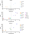
Effect of surface charge, particle size, and surface modification (PEG and ICG) on the mobility of lipid-based formulations in the vitreous based on the corresponding Dw/Dv ratios. Formulations are categorized in three graphs based on their size range: <50nm (top), 100–200 nm (middle), and >200 nm (bottom). PEGylated formulations are displayed in filled symbols, and the empty symbols represent the non-PEGylated formulations. (A) anionic, (N) neutral, (C) cationic formulation; (L) light-activated liposomes and controls; (R) rigid-membrane liposomes; (N) nanostructured lipid carriers (NLCs). See Table 1 for the detailed lipid compositions. Dv was derived from the ensemble-averaged MSD at a time scale of 1 s from at least 50 trajectories in 3 different experiments. Values for Dv (including the standard deviation), Dw, and Dw/Dv ratios are provided as Supporting Information. The Dw/Dv ratio for hexosomes is 8.7; the data is not presented in this figure due to the missing ζ potential value.
Figure 3
Figure 3
Surface plasmon resonance analysis of soft (SC) and hard (HC) corona formation on sensor-immobilized anionic liposomes with (AL1-PEG, 50 nm) and without (AL2, 50 nm) polyethylene glycol from replicate measurements with (A) thickness and (B) corresponding protein amount. Individual data points are displayed with mean and standard deviation.
Figure 4
Figure 4
Heatmap of Z-score normalized hierarchical cluster analysis with 76 differentially abundant proteins that distinguish between the hard (HC) and soft corona (SC) subsections and the porcine vitreous (source). The range is two standard deviations from the mean in both directions for relative enrichment (red) and depletion (blue) on a log2 scale. The gray color indicates that the protein was not identified in the sample replicate.

Similar articles

Cited by

References

    1. del Amo E. M.; Rimpelä A.-K.; Heikkinen E.; Kari O. K.; Ramsay E.; Lajunen T.; Schmitt M.; Pelkonen L.; Bhattacharya M.; Richardson D.; Subrizi A.; Turunen T.; Reinisalo M.; Itkonen J.; Toropainen E.; Casteleijn M.; Kidron H.; Antopolsky M.; Vellonen K.-S.; Ruponen M.; Urtti A. Pharmacokinetic Aspects of Retinal Drug Delivery. Prog. Retinal Eye Res. 2017, 57, 134–185. 10.1016/j.preteyeres.2016.12.001. - DOI - PubMed
    1. Le Goff M. M.; Bishop P. N. Adult Vitreous Structure and Postnatal Changes. Eye 2008, 22 (10), 1214–1222. 10.1038/eye.2008.21. - DOI - PubMed
    1. Käsdorf B. T.; Arends F.; Lieleg O. Diffusion Regulation in the Vitreous Humor. Biophys. J. 2015, 109 (10), 2171–2181. 10.1016/j.bpj.2015.10.002. - DOI - PMC - PubMed
    1. Pitkänen L.; Ruponen M.; Nieminen J.; Urtti A. Vitreous Is a Barrier in Nonviral Gene Transfer by Cationic Lipids and Polymers. Pharm. Res. 2003, 20 (4), 576–583. 10.1023/A:1023238530504. - DOI - PubMed
    1. Peeters L.; Sanders N. N.; Braeckmans K.; Boussery K.; Voorde J. V. de; Smedt S. C. D.; Demeester J. Vitreous: A Barrier to Nonviral Ocular Gene Therapy. Invest. Ophthalmol. Visual Sci. 2005, 46 (10), 3553–3561. 10.1167/iovs.05-0165. - DOI - PubMed

Publication types