. 2020 Jun 24;12(13):12582-12597.
doi: 10.18632/aging.103515. Epub 2020 Jun 24.

Modulation of attention networks serving reorientation in healthy aging

Affiliations

Modulation of attention networks serving reorientation in healthy aging

Yasra Arif et al. Aging (Albany NY). .

Abstract

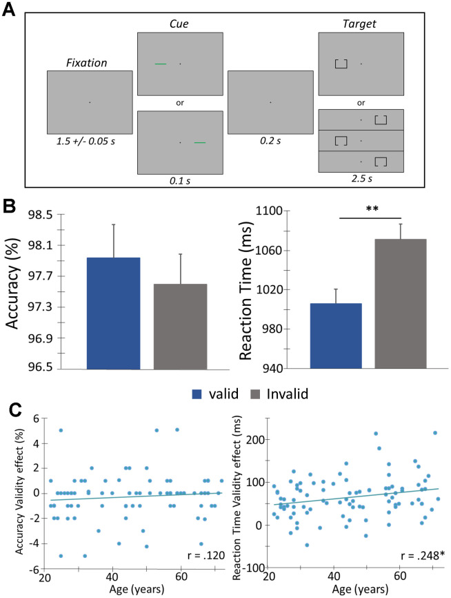
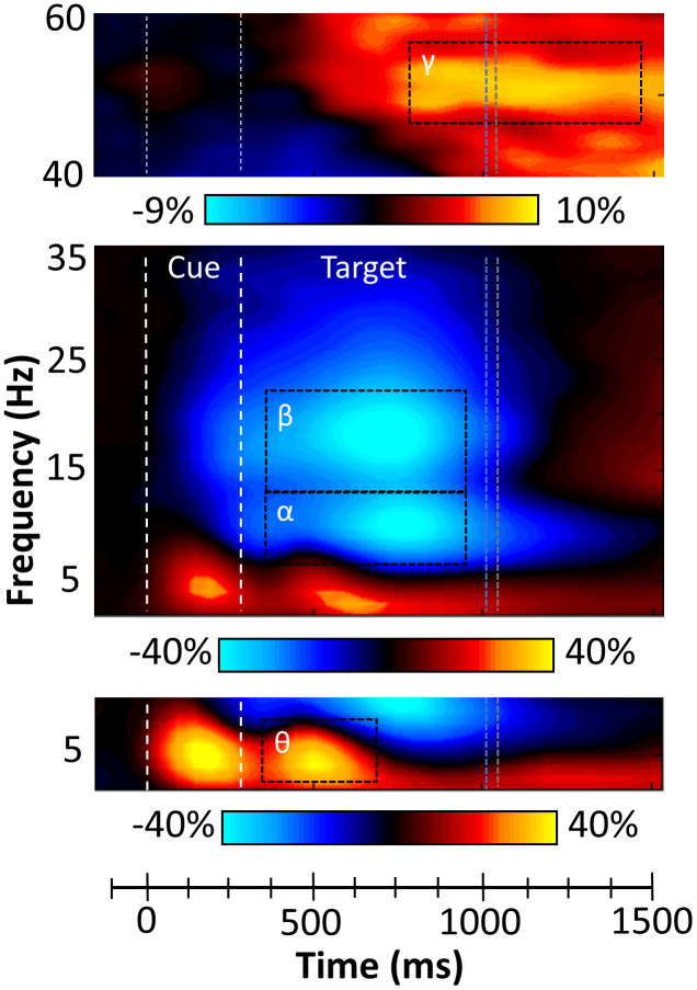
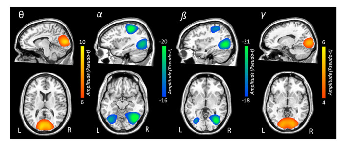
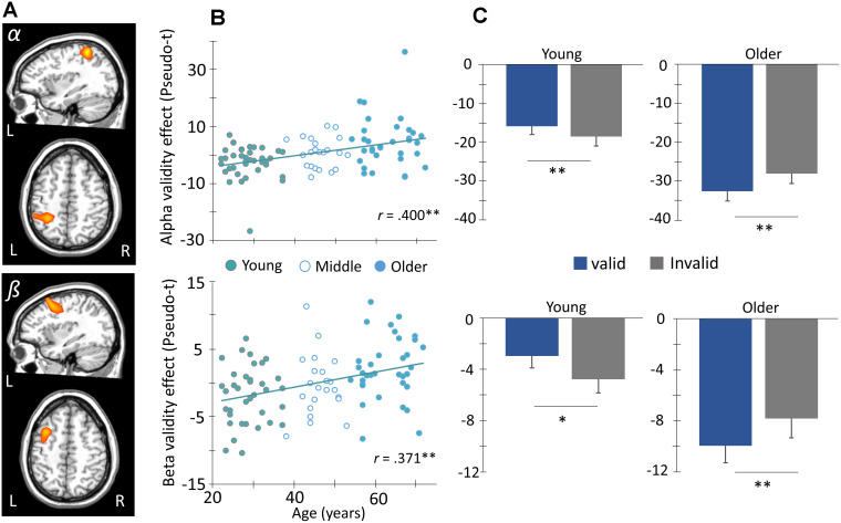
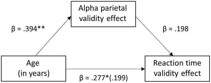
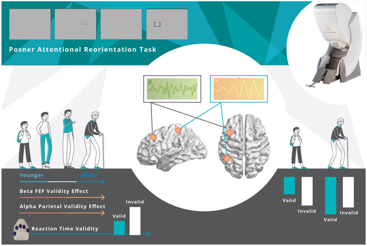
Orienting attention to behaviorally relevant stimuli is essential for everyday functioning and mainly involves activity in the dorsal and ventral frontoparietal networks. Many studies have shown declines in the speed and accuracy of attentional reallocation with advancing age, but the underlying neural dynamics remain less understood. We investigated this age-related decline using magnetoencephalography (MEG) and a Posner task in 94 healthy adults (22-72 years old). MEG data were examined in the time-frequency domain, and significant oscillatory responses were imaged using a beamformer. We found that participants responded slower when attention reallocation was needed (i.e., the validity effect) and that this effect was positively correlated with age. We also found age-related validity effects on alpha activity in the left parietal and beta in the left frontal-eye fields from 350-950 ms. Overall, stronger alpha and beta responses were observed in younger participants during attention reallocation trials, but this pattern was reversed in the older participants. Interestingly, this alpha validity effect fully mediated the relationship between age and behavioral performance. In conclusion, older adults were slower in reorienting attention and exhibited age-related alterations in alpha and beta responses within parietal and frontal regions, which may reflect increased task demands depleting their compensatory resources.

Keywords: CRUNCH; magnetoencephalography; oscillations; posner; validity effect.


Conflict of interest statement

CONFLICTS OF INTEREST: The authors of this manuscript acknowledge no conflicts of interest, financial or otherwise.

Figures

Figure 1
Figure 1
Posner cueing task and behavioral performance. (A) A central crosshair was presented for 1500 ms (± 50 ms), followed by a cue (green bar) that appeared in either the left or right hemifield for 100 ms. Target presentation (box with opening at the top or bottom) was presented 200 ms after cue offset, in either hemifield for 2500 ms. The cue was predictive of the upcoming target location 50% of the time (i.e., “valid” condition - presented on same side as the subsequent target). Participants completed 200 trials and were instructed to respond as to whether the opening was on the bottom (right index finger) or top (right middle finger) of the box. Trials were pseudorandomized and counterbalanced in regard to target validity (valid or invalid), visual hemifield (left or right), and box opening (top or bottom). (B) Behavioral metrics are displayed on the y-axis with conditions (valid or invalid) on the x-axis. Irrespective of age, participants were faster to respond and more accurate during valid compared to invalid trials. (C) Behavioral validity effect scores (invalid – valid) were computed for accuracy and reaction time and assessed as a function of age using Pearson correlations. There was a significant correlation among reaction time validity and age, such that as age increased, the difference in the reaction time between the two conditions (i.e., valid and invalid) increased. Error bars reflect the SEM. ** p < .05.
Figure 2
Figure 2
Sensor level time-frequency analysis. Grand averaged spectrograms for two sensors near parietal cortices with time (ms) displayed on the x-axis and frequency (Hz) denoted on the y-axis. Power is shown in percentage units relative to the baseline period (-600 to 0 ms), with a color scale bar beneath each spectrogram. The data per spectrogram have been averaged across all trials and participants. (Bottom) A strong increase in theta (3-7 Hz) power was observed following cue onset and during target processing (350-700 ms). (Middle) Strong decreases in alpha (8-14 Hz, 350-950 ms) and beta (14-22 Hz, 350-950 ms) power were also observed after the onset of the target. (Top) Robust increases in gamma (46-58 Hz) activity occurred during later target processing (850-1450 ms). All four oscillatory responses significantly differed from baseline activity in the same spectral band (p < .001, corrected), and these time-frequency windows have been highlighted using the black dotted line boundaries. Vertical blue and grey dotted lines represent the average reaction times for valid and invalid trials, respectively.
Figure 3
Figure 3
Grand-averaged beamformer images (pseudo-t) for each oscillatory response. In each image, data from both conditions and all participants have been grand averaged. Theta responses were strongest in bilateral medial occipital cortices. In contrast, robust decreases in alpha activity were seen in more lateral occipital cortices bilaterally and the left superior parietal lobule. Decreases in beta activity were also observed in bilateral occipital cortices and the left intraparietal sulcus. Gamma-frequency responses were strongest in the medial bilateral visual cortices.
Figure 4
Figure 4
Age modulates frontoparietal networks during attentional reorienting. (A) Whole-brain voxel-wise correlational analysis of alpha (8-14 Hz, top) and beta (14-22 Hz, bottom) validity maps (i.e., invalid – valid) and age revealed significant positive correlations between alpha and beta validity effects in left parietal cortex and frontal eye fields (FEF), respectively, and age. Images in two planes are shown for each. (B) The amplitude (pseudo-t) of the peak voxels shown in (A) were extracted and plotted as a function of age (x-axis) to identify the origin and distribution of the age-validity effect. Again, the parietal alpha data appears on the top with the frontal beta below. (C) Given this finding, the sample was stratified into age groups (i.e., younger and older) based on ± 0.5 SD of the full group’s mean age, such that subjects above 0.5 SDs were defined as the older group, and those below 0.5 SDs were defined as the younger group. This stratification can be seen in (B). The average amplitude of each conditional response (valid and invalid) is plotted to the right. Post-hoc paired t-tests were then conducted to identify the direction of the validity effect in each group. Asterisks mark significant validity effects (p < .05), with error bars reflecting the SEM. ** p < .001.
Figure 5
Figure 5
Mediation analysis of age on reaction time validity through the mediator (neural validity). There was a significant full mediation of age on reaction time validity through the mediator (i.e., alpha validity effect in the left parietal cortex), such that the increase in reaction time validity scores (i.e., cost of attention reallocation) with increased age was driven by stronger alpha desynchronizations to valid relative to invalidly-cued trials in the left parietal cortex. Each arrow is labeled with the standardized Beta coefficient values for the respective regression model. * p < .01, ** p <.005.
Figure 6
Figure 6
Overall Findings. Healthy aging modulates the behavioral and neural responses underlying attentional reorientation. The decrement in reaction time to invalid versus valid trials increased as a function of aging. Younger adults uniquely utilized parietal alpha and FEF beta activity, mainly in response to invalid trials, but this compensatory process became exhausted with increasing age.

References

    1. Corbetta M, Patel G, Shulman GL. The reorienting system of the human brain: from environment to theory of mind. Neuron. 2008; 58:306–24. 10.1016/j.neuron.2008.04.017 - DOI - PMC - PubMed
    1. Petersen SE, Posner MI. The attention system of the human brain: 20 years after. Annu Rev Neurosci. 2012; 35:73–89. 10.1146/annurev-neuro-062111-150525 - DOI - PMC - PubMed
    1. Corbetta M, Shulman GL. Control of goal-directed and stimulus-driven attention in the brain. Nat Rev Neurosci. 2002; 3:201–15. 10.1038/nrn755 - DOI - PubMed
    1. Yantis S. Control of visual attention. Attention. 1998; 1:223–256. Psychology Press/Erlbaum (UK).
    1. Wolfe JM, Butcher SJ, Lee C, Hyle M. Changing your mind: on the contributions of top-down and bottom-up guidance in visual search for feature singletons. J Exp Psychol Hum Percept Perform. 2003; 29:483–502. 10.1037/0096-1523.29.2.483 - DOI - PubMed

Publication types