Review
. 2020 Oct;75(10):2445-2476.
doi: 10.1111/all.14462.

Immunology of COVID-19: Mechanisms, clinical outcome, diagnostics, and perspectives-A report of the European Academy of Allergy and Clinical Immunology (EAACI)

Affiliations
Review

Immunology of COVID-19: Mechanisms, clinical outcome, diagnostics, and perspectives-A report of the European Academy of Allergy and Clinical Immunology (EAACI)

Milena Sokolowska et al. Allergy. 2020 Oct.

Abstract

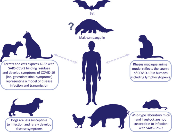
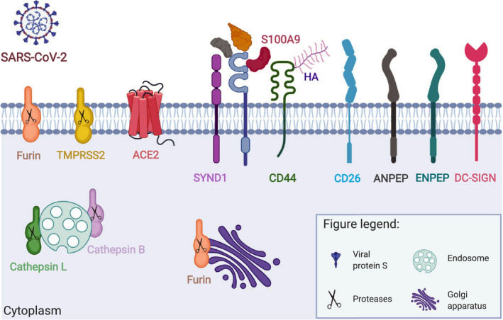
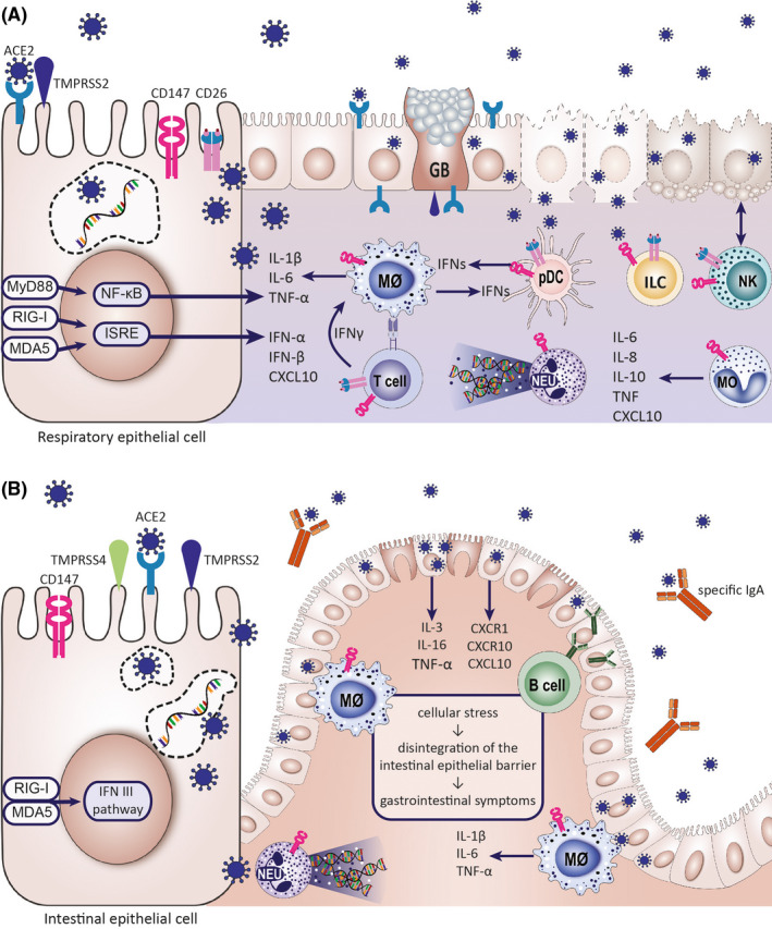
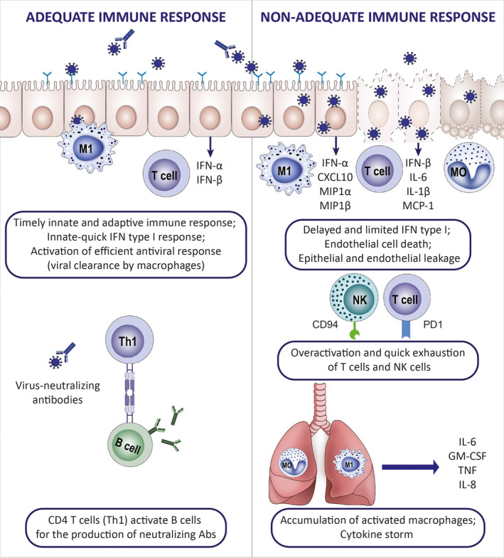
With the worldwide spread of the novel severe acute respiratory syndrome coronavirus-2 (SARS-CoV-2) resulting in declaration of a pandemic by the World Health Organization (WHO) on March 11, 2020, the SARS-CoV-2-induced coronavirus disease-19 (COVID-19) has become one of the main challenges of our times. The high infection rate and the severe disease course led to major safety and social restriction measures worldwide. There is an urgent need of unbiased expert knowledge guiding the development of efficient treatment and prevention strategies. This report summarizes current immunological data on mechanisms associated with the SARS-CoV-2 infection and COVID-19 development and progression to the most severe forms. We characterize the differences between adequate innate and adaptive immune response in mild disease and the deep immune dysfunction in the severe multiorgan disease. The similarities of the human immune response to SARS-CoV-2 and the SARS-CoV and MERS-CoV are underlined. We also summarize known and potential SARS-CoV-2 receptors on epithelial barriers, immune cells, endothelium and clinically involved organs such as lung, gut, kidney, cardiovascular, and neuronal system. Finally, we discuss the known and potential mechanisms underlying the involvement of comorbidities, gender, and age in development of COVID-19. Consequently, we highlight the knowledge gaps and urgent research requirements to provide a quick roadmap for ongoing and needed COVID-19 studies.

Keywords: COVID-19 comorbidity; COVID-19 immunity; COVID-19 multimorbidity; COVID-19 prevention; COVID-19 treatment; SARS; SARS-CoV-2 receptors.


Figures

FIGURE 1
FIGURE 1
SARS‐CoV‐2 on the animal‐human interphase. Animal models that resemble clinical and pathological features of COVID‐19 are essential to investigate pathogenesis, transmission, and therapeutic strategies. SARS‐CoV‐2 shares 96.2% of its genome sequence with the bat CoV RaTG13 posing the bat as the most probable natural host of virus origin. SARS‐CoV‐2‐related coronaviruses have been identified in Malayan pangolins, which is considered as an intermediate host between bats and humans. ACE2, a critical SARS‐CoV‐2 receptor, in wild‐type mice differs from the human one; therefore, transgenic mice models with recombinant hACE2 are necessary. To this date, Rhesus macaques, with ACE2 identical to human's, have been used to study the natural course of the disease and the effectiveness of therapeutic intervention with intravenous immunoglobulins. Ferrets and cats have been shown susceptible to SARS‐CoV‐2 infection and to develop COVID‐19 symptoms including respiratory and gastrointestinal manifestations. Limited facilities and expertise in handling nonmurine species may hamper usage of the aforementioned models. Transmission between humans and animals has not been unequivocally confirmed
FIGURE 2
FIGURE 2
Cellular distribution of confirmed and potential SARS‐CoV‐2 receptors and interaction partners. Entry of SARS‐CoV‐2 into the host cells depends on expression of i) adequate receptors and ii) cellular proteases. The two‐step infection process is mediated by the viral Spike (S) protein. Its binding to the receptor and cleavage by proteases assures virus internalization. ACE2 and TMPRSS2 are critical complex for SARS‐CoV‐2 infection. CD147 and its extracellular (Cyclophilin A, Cyclophilin B, Platelet glycoprotein VI, S100A9, Hyaluronic acid) and transmembrane (CD44, Syndecan‐1) interaction partners can be also used for SARS‐CoV‐2 entry and/or modulation of immune responses to the virus. It has been suggested for SARS‐CoV‐2 and shown in case of other Coronavidae family members that they can also exploit other cell surface receptors (CD26, ANPEP, ENPEP, DC‐SIGN) and proteases (Furin, Cathepsin L, Cathepsin) to enter human cells. CypA, Cyclophilin A; CypB, Cyclophilin B; GPVI, Platelet glycoprotein VI; HA, Hyaluronic acid; SYND1, Syndecan‐1. This figure is modified from the original publication by Radzikowska, Ding, et al, presenting the distribution of these receptors in various human tissues and immune cells in healthy children and adults, and in patients with COVID‐19 comorbidities and risk factors (ref). Created with Biorender.com
FIGURE 3
FIGURE 3
Epithelial barriers are susceptible for the SARS‐CoV‐2 infection. (A) Epithelial cells of the respiratory system are the primary site of SARS‐CoV‐2 infection. The respiratory epithelium is equipped with the receptors and other host proteins allowing viral entry: ACE2, TMPRSS2, CD147, and CD26. The highest expression of ACE2 is found in the nasopharynx. The virus was found to propagate in the lower respiratory tract as well, especially in type II alveolar cells. The effects of the virus on the respiratory epithelial barrier include cell membrane fusion and syncytium formation (which represents a mechanism of viral spread), apoptosis and virus‐mediated cell lysis leading to the loss of barrier function. Upon infection, epithelial cells release interferons, chemokines, and cytokines promoting tissue infiltration by innate immune cells, such as monocytes, NK cells, neutrophils, and, with time, inflammatory macrophages and virus‐specific lymphocytes. Immune cells express putative SARS‐CoV‐2 receptors, CD147, and CD26. (B) Gastrointestinal symptoms are seen in a substantial percentage of COVID‐19 patients. The intestinal tissue has a high expression of ACE2 receptor, TMPRSS2, and TMPRSS4 proteases. Their expression increases with intestinal epithelial cell differentiation. ACE2 expression in intestinal epithelium decreases with inflammation and shows a negative correlation with IL‐1β levels. SARS‐CoV‐2 infection results in disintegration of the intestinal epithelial barrier. Virus‐specific IgA have been found in the gastrointestinal tract. Noninfectious SARS‐CoV‐2 RNA is found in stool after negative nasal swab tests. CXCL10, C‐X‐C motif chemokine 10; CXCR1, C‐X‐C motif chemokine receptor 1; CXCR10, C‐X‐C motif chemokine receptor 10; GB, goblet cell; ILC, innate lymphoid cell; IL, interleukin; IFN, interferon; Mθ, macrophage; MO, monocyte; NEU, neutrophil; NK, natural killer cell; pDC, plasmacytoid dendritic cell; TNF, tumor necrosis factor
FIGURE 4
FIGURE 4
Immunology of adequate and nonadequate response to SARS‐CoV‐2 infection. The clinical course of the SARS‐CoV‐2 infection varies from an asymptomatic to a severe, life‐threatening syndrome. The number of asymptomatic carriers is unknown, and virus detection is often accidental. Data on the immune characteristics in this group are lacking. Patients who experience mild symptoms are characterized by a transient, slight decrease in lymphocyte counts and an increase in neutrophil counts in the peripheral blood. Viral clearance in this group is convergent in time with the specific antibody production. Delayed and limited IFN type I response in combination with the overactivation of pro‐inflammatory cytokine response has been suggested as a possible mechanistic explanation of hyperinflammatory syndrome in COVID‐19 patients presenting with severe clinical manifestations: respiratory insufficiency, kidney failure, thromboembolic, and other complications. Severe COVID‐19 is characterized by a systemic cytokine release syndrome (CRS), increased levels of LDH and CRP, hypoalbuminemia, deepening decrease in lymphocyte counts and immune exhaustion of T cells
FIGURE 5
FIGURE 5
Involvement of endothelium in COVID‐19 progression. SARS‐CoV‐2 viremia is seen approximately 1 wk after the onset of illness, accompanied by an abundance of circulating pro‐inflammatory cytokines. Endothelial cells express ACE2 receptor and can be infected by the SARS‐CoV‐2. Direct viral influence on the endothelial cells, as well as systemic inflammation (depicted by activated neutrophils and extensive NET‐osis) and cytokine storm, can lead to endotheliitis, disseminated intravascular coagulation, and coagulopathy, described in severely affected COVID‐19 patients. Activated endothelial cells upregulate the expression of adhesion molecules (P‐selectin) and coagulation factors (vWF), secrete immune mediators (CCL2, IL‐6). Monocytes respond to these by releasing tissue factor and upregulate the expression of PSGL. Simultaneously, platelet activation and aggregation occurs. Increased numbers of neutrophils and monocytes in the peripheral blood correlate with severe disease course and fatalities. CCL2, CC‐chemokine ligand; IL‐6, interleukin 6; MO, monocyte; NEU, neutrophil; NET, neutrophil extracellular traps; PLT, platelets; PSGL, P‐selectin glycoprotein ligand 1; vWF von Willebrand factor.
FIGURE 6
FIGURE 6
Age, gender, and comorbidities modify the onset and progression of COVID‐19. Epidemiological observations show clear differences in the course of SARS‐CoV‐2 infection between children and adults. It seems that children are less susceptible to the infection and develop less typical symptoms of the disease. Consequences of the infection on physiological development of children are unknown. Clinical data and age‐related rhesus macaque model of COVID‐19 reveal that obesity, diabetes, hypertension, smoking, chronic respiratory diseases, male gender, and older age are the most common risk factors for development of severe COVID‐19. Older age is associated with higher incidence of multimorbidity and state of low‐grade systemic inflammation. Immunosenescence could influence the adequacy of the host's response to the infection
FIGURE 7
FIGURE 7
Clinical stages of COVID‐19 and their virology and immunological assessment. The success of restraining SARS‐CoV‐2 transmission depends on accurate and timely diagnostics. Asymptomatic patients transmit SARS‐CoV‐2. RT‐PCR‐based test detecting the SARS‐CoV‐2 RNA in posterior conchae nasal swabs are currently the golden standard in the initial phase of the infection. Viral antigens can be detected in patients’ blood by means of ELISA tests. ELISA tests allow for detection of virus‐specific antibodies in patients’ serum. The production of specific IgM starts after about a week from infection and IgM levels decrease with the production of specific IgG (after about 2 wks from infection). Novel diagnostic and risk‐stratification strategies could include microbiome profiling and tests detecting neutralizing antibodies

Similar articles

Cited by

References

    1. Li W, Shi Z, Yu M, et al. Bats are natural reservoirs of SARS‐like coronaviruses. Science. 2005;310(5748):676‐679. - PubMed
    1. Cui J, Li F, Shi ZL. Origin and evolution of pathogenic coronaviruses. Nat Rev Microbiol. 2019;17(3):181‐192. - PMC - PubMed
    1. Zhang YZ, Holmes EC. A genomic perspective on the origin and emergence of SARS‐CoV‐2. Cell. 2020;181(2):223‐227. - PMC - PubMed
    1. Wu A, Peng Y, Huang B, et al. Genome composition and divergence of the novel coronavirus (2019‐nCoV) originating in China. Cell Host Microbe. 2020;27(3):325‐328. - PMC - PubMed
    1. Wang Q, Zhang Y, Wu L, et al. Structural and functional basis of SARS‐CoV‐2 entry by using human ACE2. Cell. 2020;181(4):894‐904.e9. - PMC - PubMed