. 2020 Aug 5;11(15):2340-2347.
doi: 10.1021/acschemneuro.0c00290. Epub 2020 Jul 14.

X-ray Crystallography Reveals Parallel and Antiparallel β-Sheet Dimers of a β-Hairpin Derived from Aβ16-36 that Assemble to Form Different Tetramers

X-ray Crystallography Reveals Parallel and Antiparallel β-Sheet Dimers of a β-Hairpin Derived from Aβ16-36 that Assemble to Form Different Tetramers

Adam G Kreutzer et al. ACS Chem Neurosci. .

Abstract

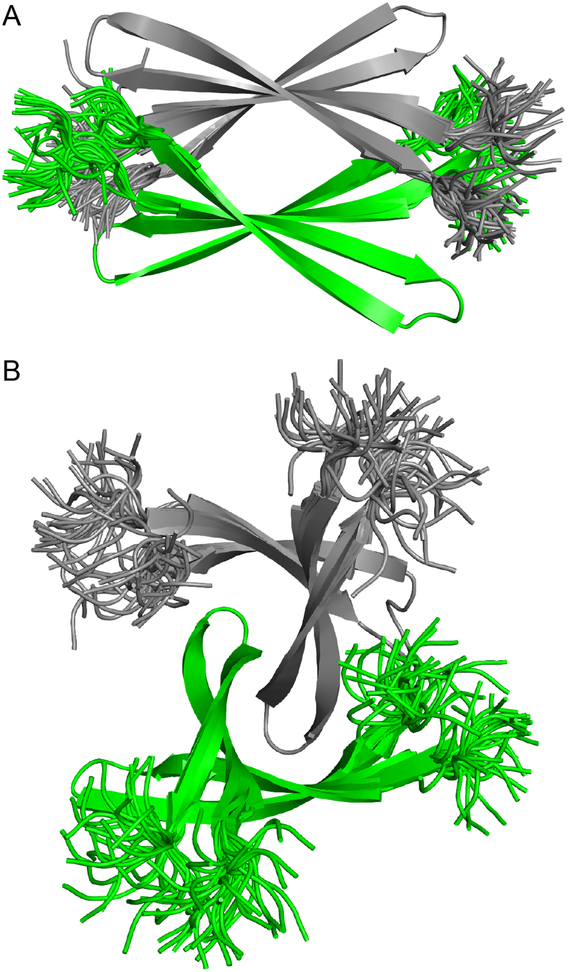
High-resolution structures of oligomers formed by the β-amyloid peptide, Aβ, are important for understanding the molecular basis of Alzheimer's disease. Dimers of Aβ are linked to the pathogenesis and progression of Alzheimer's disease, and tetramers of Aβ are neurotoxic. This paper reports the X-ray crystallographic structures of dimers and tetramers, as well as an octamer, formed by a peptide derived from the central and C-terminal regions of Aβ. In the crystal lattice, the peptide assembles to form two different dimers-an antiparallel β-sheet dimer and a parallel β-sheet dimer-that each further self-assemble to form two different tetramers-a sandwich-like tetramer and a twisted β-sheet tetramer. The structures of these dimers and tetramers derived from Aβ serve as potential models for dimers and tetramers of full-length Aβ that form in vitro and in Alzheimer's disease-afflicted brains.

Keywords: Alzheimer’s disease; Amyloid; Aβ; Crystal structure; Dimer; Oligomer; Tetramer.


Conflict of interest statement

The authors declare no competing financial interest.

Figures

Figure 1.
Figure 1.
Peptide 1. (A) Chemical structure of an Aβ16–36 β-hairpin. (B) Chemical structure of peptide 1. (C) X-ray crystallographic structure of a representative β-hairpin monomer formed by peptide 1 (PDB 6WXM). (D) Overlay of the 11 peptide 1 β-hairpins in the asymmetric unit.
Figure 2.
Figure 2.
Dimers formed by peptide 1. (A) Chemical structure (top) and X-ray crystallographic structure (bottom) of the antiparallel dimer formed by peptide 1. (B) Chemical structure (top) and X-ray crystallographic structure (bottom) of the parallel dimer formed by peptide 1.
Figure 3.
Figure 3.
X-ray crystallographic structure of the sandwich-like tetramer (dimer of antiparallel dimers) formed by peptide 1. (A) Cartoon and stick model of the sandwich-like tetramer. (B) Cartoon and sphere model of the sandwich-like tetramer. The residues that comprise the hydrophobic core are shown as spheres.
Figure 4.
Figure 4.
The twisted β-sheet tetramer formed by the parallel dimer. (A) Chemical structure. (B) X-ray crystallographic structure of the twisted β-sheet tetramer (cartoon and stick model; the side chains are omitted for clarity). (C) X-ray crystallographic structure of the twisted β-sheet tetramer (cartoon and sphere model; the residues that comprise the hydrophobic core are shown as spheres).
Figure 5.
Figure 5.
X-ray crystallographic structure of the octamer (dimer of twisted β-sheet tetramers) formed by peptide 1. (A) Cartoon and stick model of the octamer (side chains are omitted for clarity). (B) Cartoon and sphere model of the octamer illustrating the hydrophobic packing between the two twisted β-sheet tetramers. The side chains of the hydrophobic core are shown as spheres.
Figure 6.
Figure 6.
(A) The asymmetric unit of the X-ray crystallographic structure of peptide 1. (B) The crystal lattice of peptide 1, illustrating the relationship between the sandwich-like tetramer (magenta) and the twisted β-sheet tetramers that comprise the octamer (cyan). An antiparallel dimer that rests on the octamer is shown in yellow.
Figure 7.
Figure 7.
Silver-stained SDS-PAGE of recombinantly expressed Aβ40 and Aβ42 illustrating the oligomers that the peptides form in vitro. A 5-μL aliquot of each peptide concentration in a serial dilution was run on the gel.
Figure 8.
Figure 8.
Crystallographically based models of an Aβ12–40 sandwich-like tetramer (A) and an Aβ12–40 twisted β-sheet tetramer (B). Superpositions of 32 structures generated by replica-exchange molecular dynamics.
Figure 9.
Figure 9.
Representative oligomers of Aβ-derived peptides observed in our laboratory by X-ray crystallography.

Similar articles

Cited by

References

    1. Paravastu AK, Leapman RD, Yau WM, Tycko R. Molecular structural basis for polymorphism in Alzheimer’s beta-amyloid fibrils. Proc Natl Acad Sci U S A. 2008. Nov 25;105(47):18349–54. Epub 2008 Nov 17. - PMC - PubMed
    1. Petkova AT, Yau WM, Tycko R. Experimental constraints on quaternary structure in Alzheimer’s beta-amyloid fibrils. Biochemistry. 2006. Jan 17;45(2):498–512. - PMC - PubMed
    1. Lu JX, Qiang W, Yau WM, Schwieters CD, Meredith SC, Tycko R. Molecular structure of β-amyloid fibrils in Alzheimer’s disease brain tissue. Cell. 2013. Sep 12;154(6):1257–68. - PMC - PubMed
    1. Qiang W, Yau WM, Luo Y, Mattson MP, Tycko R. Antiparallel β-sheet architecture in Iowa-mutant β-amyloid fibrils. Proc Natl Acad Sci U S A. 2012. Mar 20;109(12):4443–8. Epub 2012 Mar 8. - PMC - PubMed
    1. Wälti MA, Ravotti F, Arai H, Glabe CG, Wall JS, Böckmann A, Güntert P, Meier BH, Riek R. Atomic-resolution structure of a disease-relevant Aβ(1–42) amyloid fibril. Proc Natl Acad Sci U S A. 2016. Aug 23;113(34):E4976–84. Epub 2016 Jul 28. - PMC - PubMed

Publication types