

Does methylene blue reduce adhesion during the healing process after tendon repair?
- PMID: 32584721
- PMCID: PMC7489185
- DOI: 10.5606/ehc.2020.74405
Does methylene blue reduce adhesion during the healing process after tendon repair?
Abstract
Objectives: This study aims to biomechanically and histopathologically investigate the effects of methylene blue (MB) on preventing postoperative adhesion in chickens undergoing full- thickness flexor tendon incision.
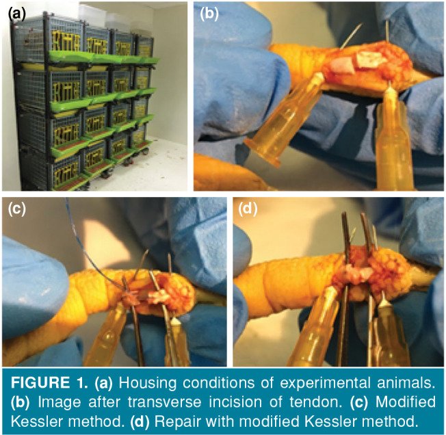
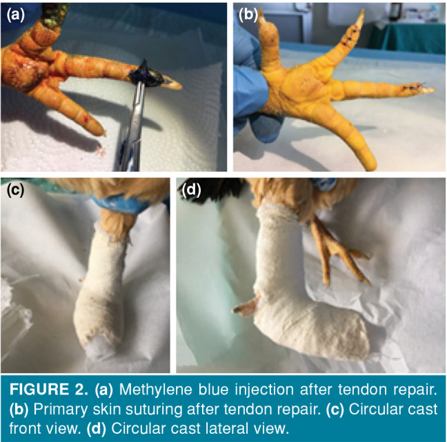
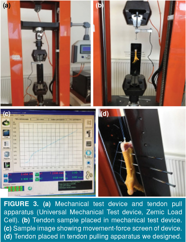
Materials and methods: This study was performed between June 2017 and June 2018 on Hubbard JA 57 type chickens (age, 6 months; weight, 2.2±0.3 kg). Sixty-four tendons were studied in 32 chickens, including 16 in the control group and 16 in the experimental group. The second and third finger flexor digitorum profundus tendons of the left foot of each chicken were repaired primarily after a full-thickness incision approximately 1 cm proximal to the distal adhesion area. In the control (n=32) and experimental groups (n=32), 0.25 mL of normal saline and 0.25 mL of 1% MB solutions were applied locally to the surgical site, respectively. The operated limb was immobilized using a circular cast. 16 chickens were randomly selected in each group and examined at the fourth week, and the remaining 16 chickens were examined at the sixth week. Thirty-two of these tendons were evaluated using the Tang histopathological adhesion classification system, and the other 32 were evaluated with a biomechanical pull system.
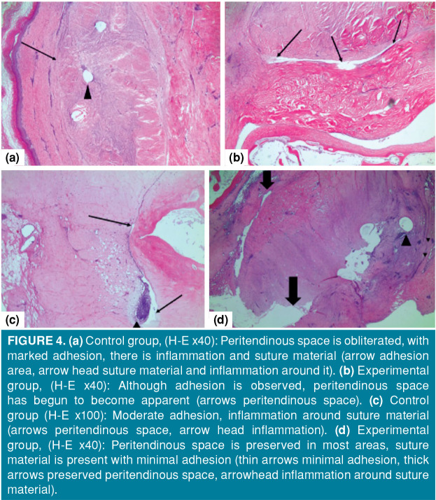
Results: Adhesion was found to be less in the experimental group compared to the control group at the end of the fourth and sixth weeks both histopathologically and biomechanically. Furthermore, adhesion was significantly less in the experimental group at the end of the sixth week compared to the fourth week both histopathologically and biomechanically.
Conclusion: Histopathological and biomechanical results show that MB, which has anti-inflammatory, antiseptic, antimicrobial and antioxidant effects, reduces adhesion during the healing process after tendon repair. We think that local MB application, particularly in surgeries performed after this type of injury, will be beneficial on early rehabilitation and functional results.
Conflict of interest statement
Figures
References
-
- Giustini M, de Leo A, Leti Acciaro A, Pajardi G, Mamo C, Voller F, et al. Incidence estimates of hand and upper extremity injuries in Italy. Ann Ist Super Sanita. 2015;51:305–312. - PubMed
-
- Clark DP, Scott RN, Anderson IW. Hand problems in an accident and emergency department. J Hand Surg Br. 1985;10:297–299. - PubMed
MeSH terms
Substances
LinkOut - more resources
Full Text Sources
Medical
Miscellaneous
