Observational Study
. 2020 Jun 25;12(12):12086-12106.
doi: 10.18632/aging.103376. Epub 2020 Jun 25.

CASC21, a FOXP1 induced long non-coding RNA, promotes colorectal cancer growth by regulating CDK6

Affiliations
Observational Study

CASC21, a FOXP1 induced long non-coding RNA, promotes colorectal cancer growth by regulating CDK6

Tao Gong et al. Aging (Albany NY). .

Abstract

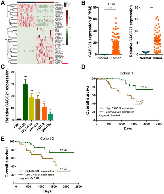
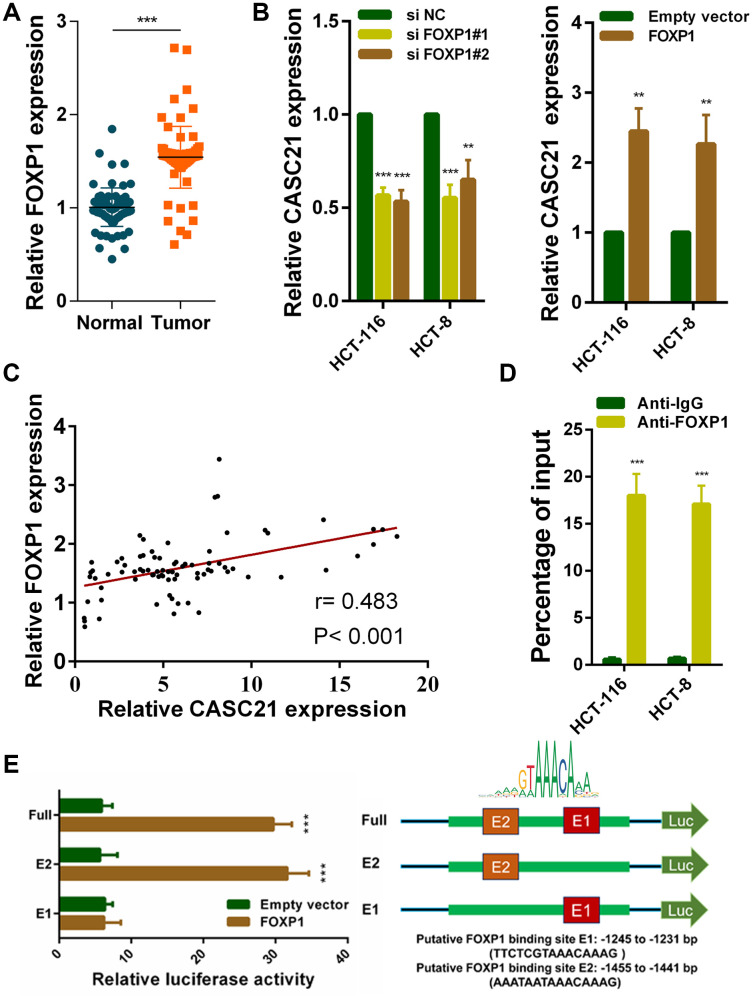
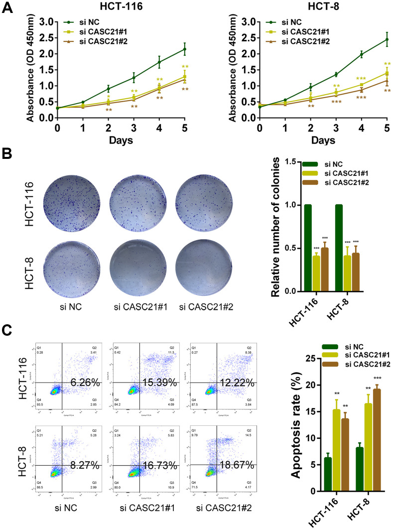
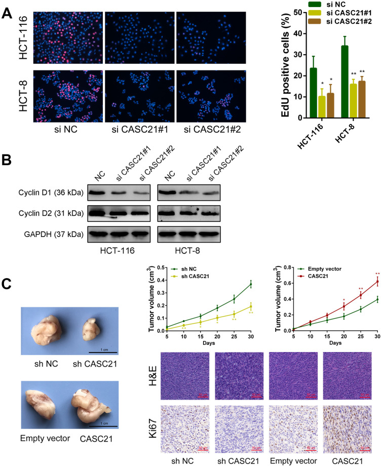
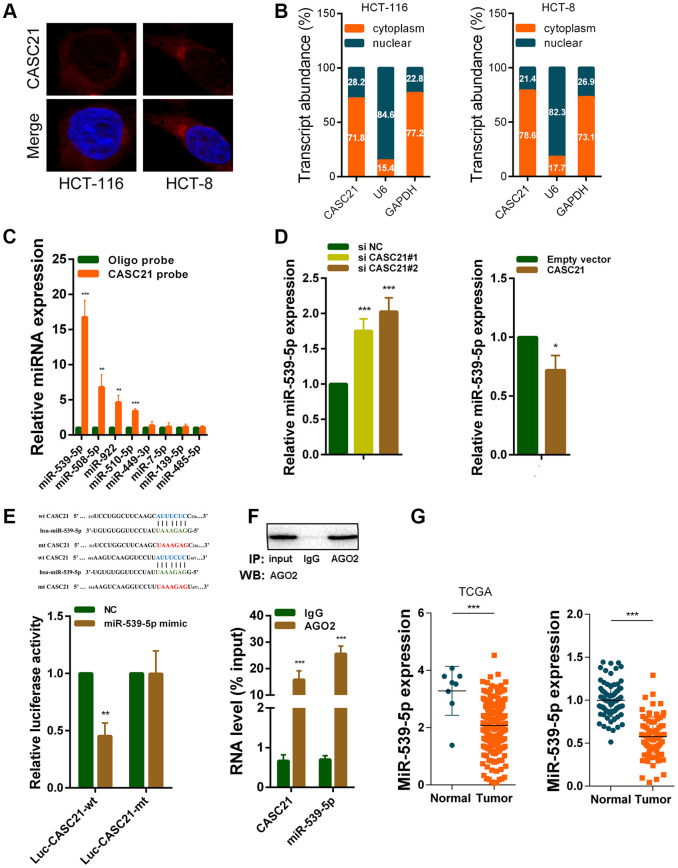
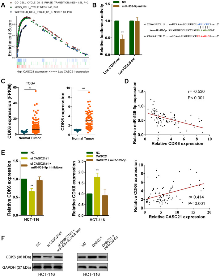
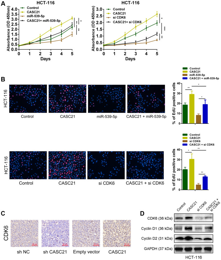
Emerging studies indicate that long non-coding RNAs (lncRNAs) play crucial roles in colorectal cancer (CRC). Here, we reported lncRNA CASC21, which is induced by FOXP1, functions as an oncogene in CRC. We systematically elucidated its clinical significance and possible molecular mechanism in CRC. LncRNA expression in CRC was analyzed by RNA-sequencing data in TCGA. The expression level of CASC21 in tissues was determined by qRT-PCR. The functions of CASC21 was investigated by in vitro and in vivo assays (CCK8 assay, colony formation assay, EdU assay, xenograft model, flow cytometry assay, immunohistochemistry (IHC) and Western blot). Chromatin immunoprecipitation (ChIP), RNA immunoprecipitation (RIP) and luciferase reporter assays were utilized to demonstrate the potential mechanisms of CASC21. CASC21 is overexpressed in CRC and high CASC21 expression is associated with poor survival. Functional experiments revealed that CASC21 promotes CRC cell growth. Mechanistically, we found that CASC21 expressed predominantly in the cytoplasm. CASC21 could interact with miR-539-5p and regulate its target CDK6. Together, our study elucidated that CASC21 acted as an oncogene in CRC, which might serve as a novel target for CRC diagnosis and therapy.

Keywords: CASC21; CDK6; colorectal cancer; lncRNA; miR-539-5p.


Conflict of interest statement

CONFLICTS OF INTEREST: We declare that we have no conflicts of interest.

Figures

Figure 1
Figure 1
CASC21 expression is elevated in CRC and high CASC21 expression predicts poor prognosis. (A) Heatmap of abnormally expressed lncRNAs in CRC in TCGA database. Red in the heat map indicates upregulation, green indicates downregulation. The red arrow denotes CASC21. (B) Left panel: expression of CASC21 in CRC generated from RNA sequencing data from TCGA database. Right panel: qRT-PCR analysis of CASC21 expression in 80 pairs of CRC and corresponding adjacent normal tissues. (C) CASC21 expression in CRC cell lines (HT-29, HCT-116, SW-620, HCT-8 and SW-480) and normal colorectal epithelial cell FHC detected by qRT-PCR. (D) Kaplan-Meier survival analysis of CRC patients’ overall survival in cohort 1 based on CASC21 expression (n= 80, P= 0.004). (E) Kaplan-Meier survival analysis of CRC patients’ overall survival in cohort 2 based on CASC21 expression (n= 64, P= 0.004). All data represent mean ± SEM (n = 3-6). *P < 0.05, **P < 0.01 and ***P < 0.001.
Figure 2
Figure 2
FOXP1 induces CASC21 high expression in CRC. (A) FOXP1 expression was detected by qRT-PCR in 80 pairs of CRC and corresponding adjacent normal tissues. (B) CASC21 expression was detected in HCT-116 and HCT-8 cells transfected with FOXP1 siRNAs or FOXP1 overexpression vector by qRT-PCR. (C) The correlation between FOXP1 and CASC21 expression analyzed in 80 paired CRC samples (n= 80, r= 0.483, P< 0.001). (D) ChIP assays were conducted to identify FOXP1 occupancy in the CASC21 promoter region. (E) Luciferase reporter assays were used to determine the FOXP1 binding sites on the CASC21 promoter region. All data represent mean ± SEM (n = 3-6). **P < 0.01 and ***P < 0.001.
Figure 3
Figure 3
CASC21 promotes CRC cells growth in vitro. (A) CCK-8 assays of HCT-116 and HCT-8 cells transfected with CASC21 siRNAs. (B) HCT-116 and HCT-8 cells transfected with CASC21 siRNAs were seeded onto 6-well plates. The number of colonies was counted on the 14th day after seeding. (C) Flow cytometric cell apoptosis assays used to assess the effect of CASC21 knockdown on cell apoptosis. All data represent mean ± SEM (n = 3-6). *P < 0.05, **P < 0.01 and ***P < 0.001.
Figure 4
Figure 4
CASC21 affects proliferation related proteins expression and promotes CRC growth in vivo. (A) EdU assays were used to determine the cell proliferate ability of CASC21 siRNAs transfected cells. (B) Cell proliferation related proteins cyclin D1, and cyclin D2, were detected by western blot after CASC21 silencing. (C) Representative images of tumors formed in nude mice from negative control vector, sh-CASC21 vector and CASC21 overexpression groups, and the tumor volume growth curves after injections in different groups. Representative images for HE-staining and Ki67 immunostaining of tumor samples from different groups. All data represent mean ± SEM (n = 3-6). *P < 0.05 and **P < 0.01.
Figure 5
Figure 5
CASC21 acts as a sponge for miR-539-5p in the cytoplasm. (A) Representative FISH images showed the location of CASC21 in HCT-116 and HCT-8 cells (red). Nuclei were stained by DAPI (blue). (B) Relative CASC21 expression levels in the nuclear and cytoplasm fractions of HCT-116 and HCT-8 cells. Nuclear controls: U6, cytosolic controls: GAPDH. (C) The relative expression of candidate microRNAs which could potentially bind to CASC21 were quantified by qRT-PCR after the biotinylated-CASC21 pull-down assays in HCT-116 cells. (D) MiR-539-5p expression was detected in HCT-116 transfected with CASC21 siRNAs or CASC21 overexpression vector by qRT-PCR. (E) Dual luciferase reporter assays of wild type and mutant type (putative binding sites for miR-539-5p were mutated) CASC21 luciferase report vectors. Up panel, sequence alignment of miR-539-5p and their predicted binding sites (green) of CASC21. Predicted micorRNA target sequence (blue) in CASC21 (Luc-CASC21-wt) and positions of mutated nucleotides (red) in CASC21 (Luc-CASC21-mt). (F) RIP assays with an anti-Ago2 antibody to assess endogenous Ago2 binding RNAs, IgG was used as the negative control. The levels of CASC21 and miR-539-5p were determined by qRT-PCR and presented as fold enrichment in Ago2 relative to input. (G) Left panel: expression of miR-539-5p in CRC generated from RNA sequencing data from TCGA database. Right panel: qRT-PCR analysis of miR-539-5p expression in 80 pairs of CRC and corresponding adjacent normal tissues. All data represent mean ± SEM (n = 3-6). *P < 0.05, **P < 0.01 and ***P < 0.001.
Figure 6
Figure 6
CASC21 regulates CDK6 expression by sponging miR-539-5p in CRC. (A) GSEA results were plotted to visualize the correlation between the expression of CASC21 and genes related to cell proliferation (KEGG_CELL_CYCLE, GO_CELL_CYCLE_G1_S_PHASE_TRANSITION and WHITFIELD_CELL_CYCLE_G1_S). (B) Dual luciferase reporter assays conducted with wild type and mutant type (putative binding sites for miR-539-5p were mutated) luciferase report vectors of CDK6 3’UTR. Right panel, sequence alignment of miR-539-5p and their predicted binding sites (green) of CDK6 3’UTR. Predicted micorRNA target sequence (blue) in CDK6 3’UTR (Luc-CDK6-wt) and positions of mutated nucleotides (red) in CDK6 3’UTR (Luc-CDK6-mt). (C) Left panel: expression of CDK6 in CRC generated from RNA sequencing data from TCGA database. Right panel: qRT-PCR analysis of CDK6 expression in 80 pairs of CRC and corresponding adjacent normal tissues. (D) Upper panel: The correlation between CDK6 and miR-539-5p in 80 paired CRC samples (n= 80, r= 0.-530, P< 0.001). Lower panel: The correlation between CDK6 and CASC21 in 80 paired CRC samples (n= 80, r= 0.414, P< 0.001). (E, F) CDK6 expression was detected by qRT-PCR or western blot in HCT-116 cells with indicated treatment. All data represent mean ± SEM (n = 3-6). **P < 0.01 and ***P < 0.001.
Figure 7
Figure 7
CASC21 promotes CRC proliferation by regulating CDK6 expression. (A) CCK-8 assays demonstrated that CASC21 promoted CRC cell growth, miR-539-5p overexpression or CDK6 knockdown could abolish growth promotion caused by CASC21. (B) EdU assays showed that miR-539-5p overexpression or CDK6 knockdown abolished the increased proliferation rates of HCT-116 cells caused by CASC21. (C) Representative images of CDK6 immunostaining of tumor samples from different groups. (D) Expression of CDK6, cyclin D1 and cyclin D2 was detected by western blot in HCT-116 cells with indicated treatment. All data represent mean ± SEM (n = 3-6). *P < 0.05, **P < 0.01 and ***P < 0.001.

Similar articles

Cited by

References

    1. Siegel RL, Miller KD, Jemal A. Cancer statistics, 2017. CA Cancer J Clin. 2017; 67:7–30. 10.3322/caac.21387 - DOI - PubMed
    1. Torre LA, Bray F, Siegel RL, Ferlay J, Lortet-Tieulent J, Jemal A. Global cancer statistics, 2012. CA Cancer J Clin. 2015; 65:87–108. 10.3322/caac.21262 - DOI - PubMed
    1. Brenner H, Kloor M, Pox CP. Colorectal cancer. Lancet. 2014; 383:1490–502. 10.1016/S0140-6736(13)61649-9 - DOI - PubMed
    1. Ferlay J, Soerjomataram I, Dikshit R, Eser S, Mathers C, Rebelo M, Parkin DM, Forman D, Bray F. Cancer incidence and mortality worldwide: sources, methods and major patterns in GLOBOCAN 2012. Int J Cancer. 2015; 136:E359–86. 10.1002/ijc.29210 - DOI - PubMed
    1. Atkinson SR, Marguerat S, Bähler J. Exploring long non-coding RNAs through sequencing. Semin Cell Dev Biol. 2012; 23:200–05. 10.1016/j.semcdb.2011.12.003 - DOI - PubMed

Publication types

MeSH terms