. 2020 Jun 25;12(12):11165-11184.
doi: 10.18632/aging.103567. Epub 2020 Jun 25.

SIRT6 mono-ADP ribosylates KDM2A to locally increase H3K36me2 at DNA damage sites to inhibit transcription and promote repair

Affiliations

SIRT6 mono-ADP ribosylates KDM2A to locally increase H3K36me2 at DNA damage sites to inhibit transcription and promote repair

Sarallah Rezazadeh et al. Aging (Albany NY). .

Abstract

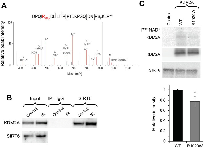
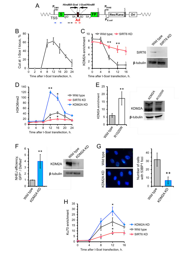
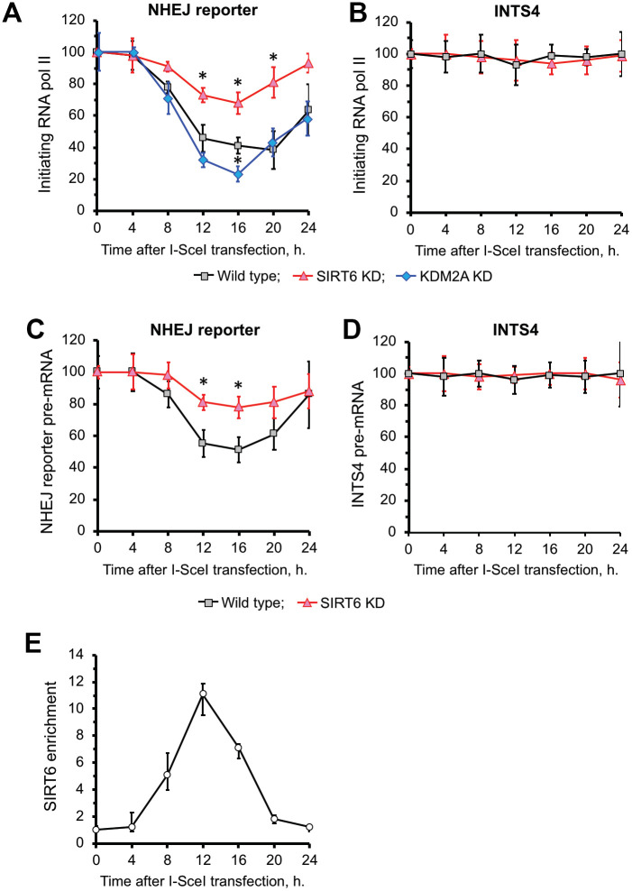
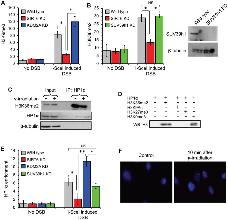
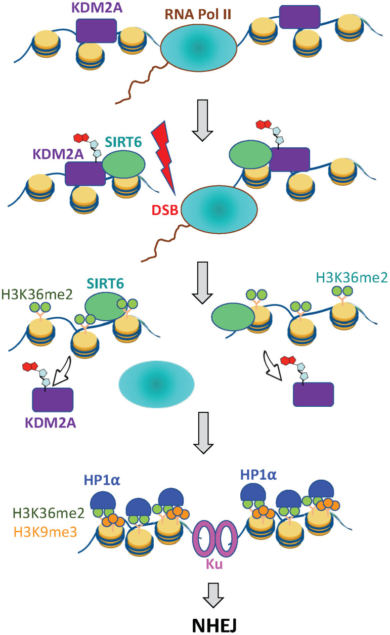
When transcribed DNA is damaged, the transcription and DNA repair machineries must interact to ensure successful DNA repair. The mechanisms of this interaction in the context of chromatin are still being elucidated. Here we show that the SIRT6 protein enhances non-homologous end joining (NHEJ) DNA repair by transiently repressing transcription. Specifically, SIRT6 mono-ADP ribosylates the lysine demethylase JHDM1A/KDM2A leading to rapid displacement of KDM2A from chromatin, resulting in increased H3K36me2 levels. Furthermore, we found that through HP1α binding, H3K36me2 promotes subsequent H3K9 tri-methylation. This results in transient suppression of transcription initiation by RNA polymerase II and recruitment of NHEJ factors to DNA double-stranded breaks (DSBs). These data reveal a mechanism where SIRT6 mediates a crosstalk between transcription and DNA repair machineries to promote DNA repair. SIRT6 functions in multiple pathways related to aging, and its novel function coordinating DNA repair and transcription is yet another way by which SIRT6 promotes genome stability and longevity.

Keywords: DNA repair; SIRT6; genome stability; longevity; transcription.


Conflict of interest statement

CONFLICTS OF INTEREST: The authors declare no conflicts of interest.

Figures

Figure 1
Figure 1
SIRT6 mono-ADP ribosylates KDM2A on R1019/R1020 (mouse/human). (A) Mass spec analysis of mouse KDM2A mono-ADP ribosylation. MS2 fragment spectrum of a KDM2A peptide obtained from mouse embryonic fibroblast cells supporting R1019 modified by a ribose-phosphate group indicating mono-ADP ribosylation (MAR). Pictured is an assignment showing ribose-phosphate modified R1019. A full compiled report downloaded from Protein Prospector of the MS2 data for this peptide can be found in the Supplementary Data (Supplementary Data 1). (B) SIRT6 interacts with KDM2A. coimmunoprecipitation of KDM2A was observed with antibodies directed against SIRT6 before and after irradiation (IR) (the experiment was repeated three times). (C) SIRT6 mono-ADP ribosylates KDM2A in vitro. KDM2A R1020W mutation significantly decreases SIRT6 mono-ADP ribosylation signal. Top panel. SIRT6 protein was incubated with purified human wild type flag-KDM2A or mutant R1020W proteins in the presence of radiolabeled NAD+. Mono-ADP ribosylation was detected by transfer of the radiolabel to the substrate. Lower panel. Quantification of upper gel. Graph represents mean ± SD of three experiments. *p > 0.05.
Figure 2
Figure 2
SIRT6 mediates KDM2A displacement from chromatin to enhance NHEJ. (A) Schematic representation of the GFP-based NHEJ reporter, which represents an RNA Pol II-transcribed gene. DSBs are introduced by transient expression of I-SceI enzyme. The positions of the primer used in this study are indicated by small arrows below: primers for quantification of the preRNA (small black arrows); quantification of DNA DSBs (small red arrows), chromatin IP (blue arrows). (B) Quantification of the cutting efficiency at different time points after transfection with I-SceI vector. DSBs were quantified as a ratio between the product obtained with primers amplifying across the break (red arrows) to a PCR product from a gene away from DSB site (Supplementary Figure 2). The experiment was repeated three times. (C) KDM2A dissociation from broken chromatin is SIRT6-dependent. Time-course chromatin-IP was performed using antibody against endogenous KDM2A in human skin fibroblasts harboring chromosomally-integrated NHEJ GFP reporter with I-SceI sites. Western blot shows depletion of SIRT6. (D) H3K36me2 accumulation post DSB induction is SIRT6-dependent. ChIP analysis of H3K36me2, at different time points post transfection with I-SceI vector using antibody specific for H3K36me2. qPCR was performed using primers positioned 50 bp downstream of TSS. (E) Non-ribosylatable KDM2A R1020W mutant bids to DSBs more efficiently than the wild type protein. Human skin fibroblasts carrying I-SceI reporter cassette were transfected with wild type or mutant (R1020W) KDM2A and I-SceI encoding vectors. At 12 hours post transfection cells were harvested followed by ChIP-qPCR with antibodies against FLAG. Western blot showing the levels of wild type and mutant FLAG-KDM2A proteins in cells after transfection. (F) Cells depleted of KDM2A have enhanced NHEJ repair efficiency. Cells were treated with siRNA to KDM2A four days before transfection with I-SceI. NHEJ efficiency was measured by reactivation of the GFP reporter normalized to transfection efficiency (DsRed) 48 h after I-SceI transfection. Western blot shows KDM2A depletion. (G) Downregulation of KDM2A reduces the number of 53BP1 foci. KDM2A knocked down cells were grown to confluency and fixed for IF experiment using antibody against 53BP1. On the right; quantification of the IF image obtained from 100 nuclei. All experiments were repeated at least three times. (H) Ku70 recruitment to DSB site requires SIRT6, and is counteracted by KDM2A. Time course of ChIP experiment using antibody against Ku70. The experiment was repeated three times. *p < 0.05; **p <0.01.
Figure 3
Figure 3
SIRT6 is required for inhibition of transcription upon induction of DNA DSB. (A) SIRT6 is required for RNA Pol II displacement upon DSB. Time course ChIP analysis of initiating (S5 phosphorylated) RNA Pol II levels on the TSS region of the GFP in NHEJ reporter upon DSB induction. (B) Time course ChIP analysis of initiating RNA Pol II levels on the TSS region of INTS4 gene, which does not undergo a DSB (negative control to A). (C) qRT-PCR analysis of GFP premature RNA expression at different time points after transfection with I-SceI vector. (D) qRT-PCR analysis of INTS4 premature RNA expression (control for C). (E) SIRT6 recruitment to DSB peaks within 12 h post DSB induction. Chromatin IP of endogenous SIRT6 in skin fibroblasts. Chromatin samples were analyzed by qPCR using primers flanking I-SceI cut sites with antibodies against SIRT6 or IgG. The positions of primers in NHEJ reporter and INTS4 gene are shown in Figures 2A and Supplementary Figure 2. The experiments were repeated three times. *p < 0.05.
Figure 4
Figure 4
H3K36me2 recruits HP1α and promotes deposition of H3K9me3 at DSB locus in SIRT6-dependent manner. (A) SIRT6, but not KDM2A, is required for deposition of H3K9me3 at DSB locus. ChIP assay was performed 12 h post transfection with I-SceI vector using antibody against H3K9me3. (B) SIRT6, but not SUV39h1, is required for accumulation of H3K36me2 at DSB locus. Western blot shows depletion of SUV39h1. (C) HP1α binds H3K36me2 in vivo. Human skin fibroblast cells were irradiated with 3 Gy of γ-irradiation. Cell lysate was immunoprecipitated within 10 minutes after irradiation with antibody against HP1α and blotted against H3K36me2 (Top panel), HP1α (middle) and β-tubulin (lower panel). (D) HP1α binds H3K36me2 in vitro. Mono nucleosomes carrying either H3K36me2, H3K27me3, H3K9me3 or H3K9Ac groups were incubated with HP1α and immunoprecipitated with antibody against HP1α and blotted with H3 antibodies. (E) SIRT6, but not SUV39h1, is required for HP1α recruitment to DSB locus. (F) HP1α interacts with 53BP1 after γ-irradiation. Proximity ligation assay (PLA) was performed using antibodies against HP1α and 53BP1 and the PLA signal was detected within 10 minutes after irradiation. NS; not significant. All experiments were repeated three times. *p < 0.05; **p <0.01.
Figure 5
Figure 5
SIRT6 prevents collision between transcription and DSB repair machineries. Under basal conditions KDM2A demethylates H3K36 at transcription start site. Upon DNA damage SIRT6, is recruited to the DSB locus and mono-ADP ribosylates KDM2A leading to its displacement from chromatin. This leads to accumulation of H3K36me2 marks around the DSB site, which recruits HP1α and promotes deposition of H3K9me3 mark leading to local chromatin compaction. As a result, transcription is paused and DNA repair by NHEJ ensues.

Similar articles

Cited by

References

    1. Ciccia A, Elledge SJ. The DNA damage response: making it safe to play with knives. Mol Cell. 2010; 40:179–204. 10.1016/j.molcel.2010.09.019 - DOI - PMC - PubMed
    1. Bekker-Jensen S, Lukas C, Kitagawa R, Melander F, Kastan MB, Bartek J, Lukas J. Spatial organization of the mammalian genome surveillance machinery in response to DNA strand breaks. J Cell Biol. 2006; 173:195–206. 10.1083/jcb.200510130 - DOI - PMC - PubMed
    1. Ui A, Nagaura Y, Yasui A. Transcriptional elongation factor ENL phosphorylated by ATM recruits polycomb and switches off transcription for DSB repair. Mol Cell. 2015; 58:468–82. 10.1016/j.molcel.2015.03.023 - DOI - PubMed
    1. Kakarougkas A, Ismail A, Chambers AL, Riballo E, Herbert AD, Künzel J, Löbrich M, Jeggo PA, Downs JA. Requirement for PBAF in transcriptional repression and repair at DNA breaks in actively transcribed regions of chromatin. Mol Cell. 2014; 55:723–32. 10.1016/j.molcel.2014.06.028 - DOI - PMC - PubMed
    1. Miller KM, Tjeertes JV, Coates J, Legube G, Polo SE, Britton S, Jackson SP. Human HDAC1 and HDAC2 function in the DNA-damage response to promote DNA nonhomologous end-joining. Nat Struct Mol Biol. 2010; 17:1144–51. 10.1038/nsmb.1899 - DOI - PMC - PubMed

Publication types