. 2020 Sep 1;130(9):4999-5010.
doi: 10.1172/JCI136621.

12(S)-HETE mediates diabetes-induced endothelial dysfunction by activating intracellular endothelial cell TRPV1

Affiliations

12(S)-HETE mediates diabetes-induced endothelial dysfunction by activating intracellular endothelial cell TRPV1

Mandy Otto et al. J Clin Invest. .

Abstract

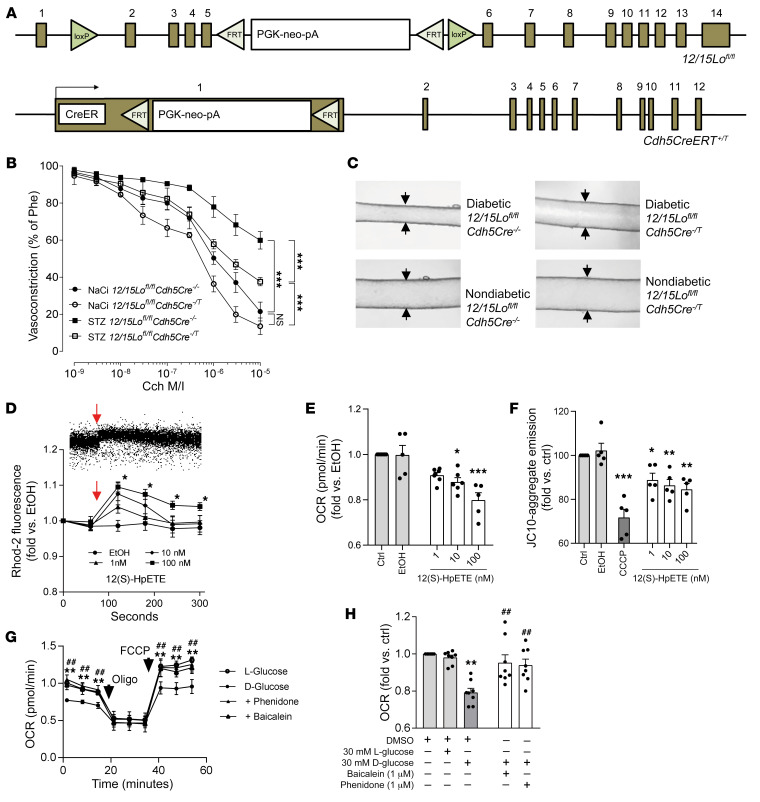
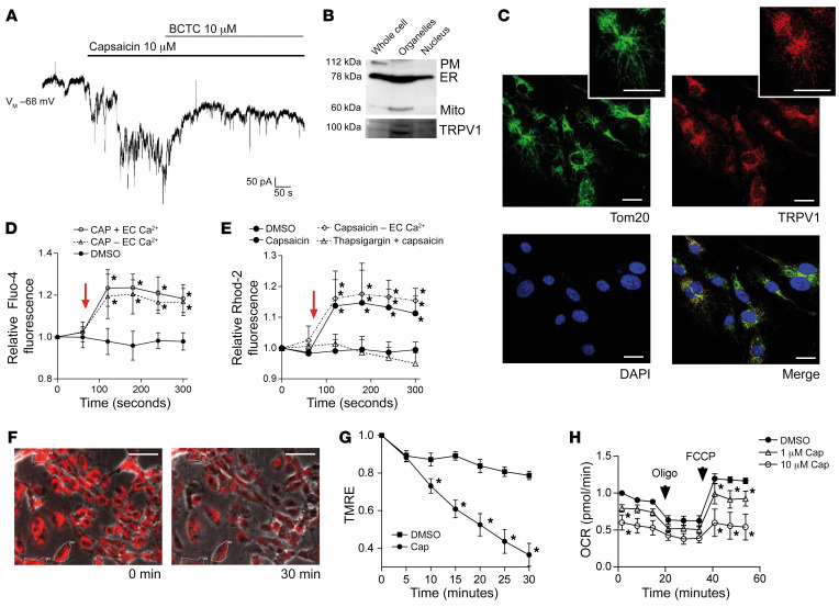
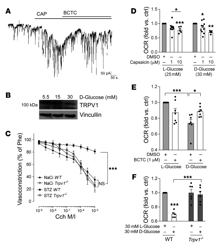
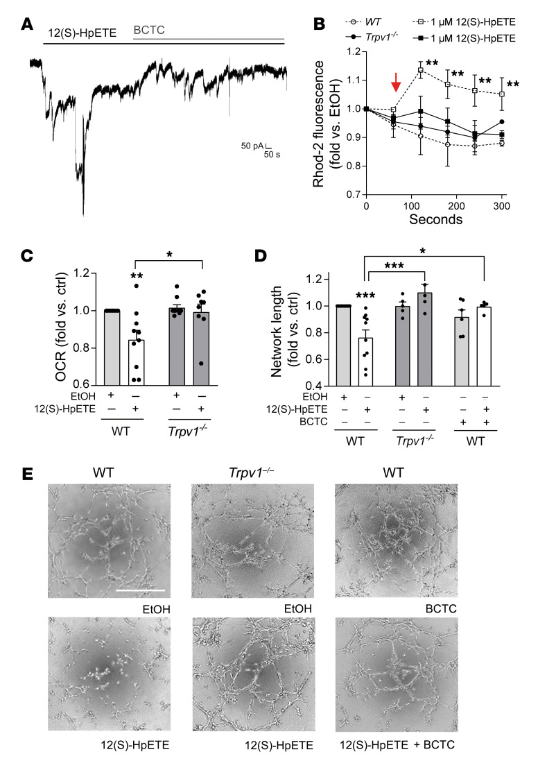
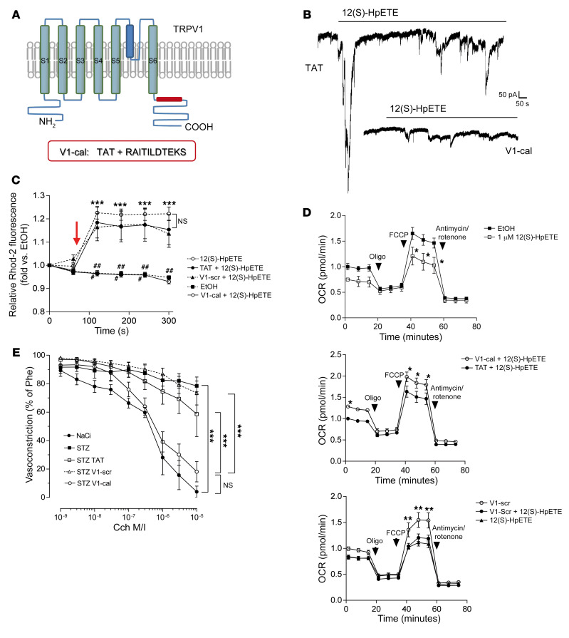
Patients with diabetes develop endothelial dysfunction shortly after diabetes onset that progresses to vascular disease underlying the majority of diabetes-associated comorbidities. Increased lipid peroxidation, mitochondrial calcium overload, and mitochondrial dysfunction are characteristics of dysfunctional endothelial cells in diabetic patients. We here identified that targeting the lipid peroxidation product 12(S)-hydroxyeicosatetraenoic acid-induced [12(S)-HETE-induced] activation of the intracellularly located cation channel transient receptor potential vanilloid 1 (TRPV1) in endothelial cells is a means to causally control early-stage vascular disease in type I diabetic mice. Mice with an inducible, endothelium-specific 12/15-lipoxygenase (12/15Lo) knockout were protected similarly to TRPV1-knockout mice from type 1 diabetes-induced endothelial dysfunction and impaired vascular regeneration following arterial injury. Both 12(S)-HETE in concentrations found in diabetic patients and TRPV1 agonists triggered mitochondrial calcium influx and mitochondrial dysfunction in endothelial cells, and 12(S)-HETE effects were absent in endothelial cells from TRPV1-knockout mice. As a therapeutic consequence, we found that a peptide targeting 12(S)-HETE-induced TRPV1 interaction at the TRPV1 TRP box ameliorated diabetes-induced endothelial dysfunction and augmented vascular regeneration in diabetic mice. Our findings suggest that pharmacological targeting of increased endothelial lipid peroxidation can attenuate diabetes-induced comorbidities related to vascular disease.

Keywords: Diabetes; Eicosanoids; Vascular Biology; endothelial cells.


Conflict of interest statement

Conflict of interest: ERG holds a patent on “Peptide modulators of specific calcineurin protein-protein interactions” (US Application Serial No. 16/082,313) that includes the V1-cal peptide.

Figures

Figure 1
Figure 1. 12(S)-HETE mediates diabetes-induced endothelial and mitochondrial dysfunction.
(A) Mice used to generate endothelium-specific 12/15Lo knockout mice. (B) Carbamoylcholine (Cch) induced vasorelaxation in murine mesenteric resistance arteries preconstricted with 10–5 M phenylephrine (Phe) in 12/15Lofl/fl mice positive (Cre–/T) or negative (Cre–/–) for the Cre transgene. Type 1 diabetes was induced by injection of streptozotocin (STZ), dissolved in sodium citrate (NaCi) serving as vehicle control in nondiabetic mice. ***P < 0.001 vs. as indicated, 2-way ANOVA/Bonferroni. n = 5 mice per group. M/I, moles per liter. (C) Representative pictures of Cch-induced vasorelaxation at 10–5 M. (D) Mitochondrial calcium influx over time detected as fluorescence intensity changes by flow cytometry in rhodamine-2–loaded (Rhod-2–loaded) human endothelial cells stimulated with 12(S)-HpETE, with time of addition indicated by arrows. Upper panel shows a representative flow cytometry dot plot, lower panel the quantitative summary of n = 6 independent experiments. Ethanol (EtOH) served as vehicle control. *P < 0.05 vs. mean baseline, 2-way ANOVA/Bonferroni. (E) Mitochondrial respiration (oxygen consumption rate, OCR) measured by a Seahorse extracellular flux analyzer. *P < 0.05, ***P < 0.001 vs. EtOH, 1-way ANOVA/Bonferroni, n = 5–7 experiments per group. (F) Decline of mitochondrial membrane potential detected by flow cytometry as loss of red fluorescence. Two micromolar CCCP was used as positive control. *P < 0.05, **P < 0.01, ***P < 0.001 vs. EtOH, 1-way ANOVA/Bonferroni, n = 5–7 independent experiments. (G) Extracellular flux analysis of high-glucose-exposed human endothelial cells. Inhibition of 12LOX by baicalein or phenidone improves mitochondrial OCR. Maximum mitochondrial respiration was assessed after addition of oligomycin (Oligo) and FCCP, indicated by arrows. **P < 0.01 baicalein, ##P < 0.01 phenidone vs. d-glucose only, 2-way ANOVA/Bonferroni. Pooled data from n = 5–8 independent experiments with each variant each analyzed in triplicate. (H) Summary of basal respiration data. **P < 0.01 vs. l-glucose, ##P < 0.01 vs. d-glucose, 1-way ANOVA/Bonferroni, n = 8 independent experiments. All graphs show mean ± SEM.
Figure 2
Figure 2. TRPV1, a receptor activated by 12(S)-HpETE, is present and functional in human endothelial cells, located intracellularly, and a mediator of mitochondrial dysfunction.
(A) Representative patch clamp experiment of human endothelial cell exposure to 10 μM capsaicin shows currents induced by TRPV1 activation that are antagonized by 10 μM of the TRPV1 antagonist BCTC. Representative patch of n = 15 independent experiments. (B) Cell fractionation reveals that TRPV1 is enriched in fractions containing ER (here identified by GRP78 expression) and mitochondria (Mito, ATP5a). PM, plasma membrane, Na+/K+-ATPase. (C) Immunohistochemistry showing that TRPV1 (red) colocalizes with Tom20 (green) in human endothelial cells. Cell nuclei are stained with DAPI. Scale bars: 20 μm. (D and E) Ten micromolar capsaicin (time point of addition indicated by arrow) induces increase in cytosolic calcium in Fluo-4–loaded HUVECs and increase in mitochondrial calcium in Rhod-2–loaded HUVECs independent of the presence (+ EC Ca2+) or absence (– EC Ca2+) of extracellular calcium but dependent on calcium in the ER since effects were absent after thapsigargin-induced ER depletion. *P < 0.01 vs. baseline fluorescence, 2-way ANOVA/Bonferroni, n = 6 independent experiments. (F) One micromolar capsaicin induces decline in mitochondrial membrane potential indicated as decline in fluorescence intensity of TMRE-loaded human endothelial cells during live-cell imaging. *P < 0.05 vs. baseline fluorescence intensity, 2-way ANOVA/Bonferroni, mean of at least 5 cells per high-power field in n = 5 independent experiments. Scale bars: 50 μm. (G) Prestimulation of human endothelial cells with 1 μM or 10 μM capsaicin reduces mitochondrial OCR. Maximum mitochondrial respiration was assessed after addition of oligomycin and FCCP, indicated by arrows. *P < 0.05 vs. baseline, 2-way ANOVA/Bonferroni. Pooled data from n = 5 independent experiments with each variant analyzed in triplicate. All graphs show mean ± SEM.
Figure 3
Figure 3. TRPV1 presence and function are unaltered under high-glucose conditions and TRPV1 deficiency protects against diabetes-induced endothelial dysfunction.
(A) Patch clamp experiments of human endothelial cells cultured under high levels of d-glucose (30 mM) show robust currents induced by 10 μM of capsaicin (CAP) that are antagonized by 10 μM of the TRPV1 antagonist BCTC. Representative result out of n = 10 independent experiments. (B) Representative Western blot of human endothelial cells cultured under high-glucose conditions for 24–96 hours (here 48 hours) shows that TRPV1 expression is not altered by high glucose. Twenty-five or 10 mM d-glucose was added to 5.5 mM d-glucose in media. Twenty-five or 15 mM l-glucose was added for osmotic control. (C) Cch-induced vasorelaxation in murine mesenteric resistance arteries preconstricted with 10–5 M phenylephrine shows that TRPV1-knockout (Trpv1–/–) mice are protected from diabetes-induced endothelial dysfunction. STZ, streptozotocin; NaCi, sodium citrate serving as vehicle control. ***P < 0.001 vs. as indicated, 2-way ANOVA/Bonferroni. Means were generated for each individual animal by analysis of 2 arteries, n = 10 animals per group. (D) Capsaicin reduces mitochondrial OCR in a comparable fashion in endothelial cells exposed to high levels of l- or d-glucose. *P < 0.05, **P < 0.01, ***P < 0.001 vs. DMSO or as indicated, 1-way ANOVA/Bonferroni, summary of n = 5–10 independent experiments. (E) Impaired mitochondrial respiration under high–d-glucose conditions (30 mM) in human endothelial cells is improved by coincubation with the TRPV1 antagonist BCTC. *P < 0.05, ***P < 0.001 vs. as indicated, 1-way ANOVA/Bonferroni, n = 7 independent experiments. (F) High levels of d-glucose impair mitochondrial respiration in endothelial cells isolated from WT but not Trpv1–/– mice. Endothelial cells were analyzed from n = 5–6 independent isolations. ***P < 0.001 vs. l-glucose or as indicated. All graphs show mean ± SEM.
Figure 4
Figure 4. 12(S)-HpETE effects on mitochondrial and endothelial function are mediated by TRPV1.
(A) Patch clamp experiments with human endothelial cells show that 700 nM 12(S)-HpETE induces currents comparable to those induced by 10 μM capsaicin and that these currents are antagonized by 10 μM of the TRPV1 antagonist BCTC. (B) Mitochondrial calcium increase induced by 1 μM 12(S)-HpETE added as indicated by the arrow and assessed as increase in fluorescence intensity of Rhod-2–loaded murine endothelial cells isolated from WT (dashed lines) versus Trpv1–/– mice. **P < 0.01 vs. baseline, 2-way ANOVA/Bonferroni, summary of n = 5 independent experiments. (C) Mitochondrial OCR is reduced by 100 nM 12(S)-HpETE in endothelial cells from WT but not Trpv1–/– mice. *P < 0.05, **P < 0.01 vs. EtOH or as indicated, 1-way ANOVA/Bonferroni, n = 5–10 independent experiments. (D and E) Effects of 12(S)-HpETE on endothelial capillary-like tube formation on Matrigel. Effects of 100 nM 12(S)-HpETE are absent in murine endothelial cells isolated from Trpv1–/– mice and endothelial cells isolated from WT mice in the presence of 10 μM of the TRPV1 antagonist BCTC. *P <0.05, ***P < 0.001 vs. EtOH or as indicated, 1-way ANOVA/Bonferroni, n = 5–10 independent experiments. Scale bar: 500 μm. All data are presented as mean ± SEM.
Figure 5
Figure 5. Abrogation of the 12(S)-HpETE/TRPV1 interaction protects against diabetes-induced endothelial dysfunction.
(A) Schematic of TRPV1 corresponding to amino acids 701–711 within the TRP box at the C-terminus of human TRPV1 linked to a TAT linker protein for intracellular entry. (B) Patch clamp experiments show that 700 nM 12(S)-HpETE evokes currents in endothelial cells loaded with 1 μM TAT linker, but not 1 μM V1-cal. Representative patch results from n = 10 independent experiments. (C) 12(S)-HpETE–induced mitochondrial calcium changes detected by flow cytometry [time point of addition of 12(S)-HpETE is indicated by the arrow] in human endothelial cells preincubated with either 1 μM V1-cal or 1 μM of a scrambled version of V1-cal (V1-scr) or TAT linker protein serving as controls. ***P < 0.001 vs. corresponding time point in V1-cal or EtOH, #P < 0.05, ##P < 0.01, ###P < 0.001 vs. TAT or V1-scr, 2-way ANOVA/Bonferroni, n = 5 independent experiments. (D) Assessment of mitochondrial function to evaluate effects of 1 μM V1-cal versus 1 μM TAT linker only (top 2 panels) or 1 μM V1-scr (bottom panel) on mitochondrial OCR induced by 1 μM 12(S)-HpETE. After 3 measurements, oligomycin was added before FCCP used to induce maximum respiration. *P < 0.05, **P < 0.01, 2-way ANOVA/Bonferroni. Variants were analyzed in triplicate on Seahorse miniplates, and n = 5 independent experiments were performed. All graphs show mean ± SEM. (E) Cch-induced vasorelaxation shows that repetitive intravenous application on 4 consecutive days of the V1-cal peptide (1 mg/kg/d), but not of the V1-scr peptide or the TAT linker protein only, to diabetic WT mice reduced endothelial dysfunction induced by diabetes (STZ). Sodium citrate (NaCi) was used as vehicle control. ***P < 0.001 vs. as indicated, 2-way ANOVA/Bonferroni. Means were generated for each individual animal by analysis of 2 arteries; n = 5 animals per group. Graph shows mean ± SEM.

Similar articles

Cited by

References

    1. Patterson CC, et al. Trends and cyclical variation in the incidence of childhood type 1 diabetes in 26 European centres in the 25 year period 1989-2013: a multicentre prospective registration study. Diabetologia. 2019;62(3):408–417. doi: 10.1007/s00125-018-4763-3. - DOI - PubMed
    1. Forbes JM, Cooper ME. Mechanisms of diabetic complications. Physiol Rev. 2013;93(1):137–188. doi: 10.1152/physrev.00045.2011. - DOI - PubMed
    1. Nascimento AMMAD, Sequeira IJ, Vasconcelos DF, Gandolfi L, Pratesi R, Nóbrega YKM. Endothelial dysfunction in children with type 1 diabetes mellitus. Arch Endocrinol Metab. 2017;61(5):476–483. doi: 10.1590/2359-3997000000271. - DOI - PMC - PubMed
    1. Kitta Y, et al. Persistent impairment of endothelial vasomotor function has a negative impact on outcome in patients with coronary artery disease. J Am Coll Cardiol. 2009;53(4):323–330. doi: 10.1016/j.jacc.2008.08.074. - DOI - PubMed
    1. Shenouda SM, et al. Altered mitochondrial dynamics contributes to endothelial dysfunction in diabetes mellitus. Circulation. 2011;124(4):444–453. doi: 10.1161/CIRCULATIONAHA.110.014506. - DOI - PMC - PubMed

Publication types

MeSH terms

Substances