. 2020 Sep;58(1):e108.
doi: 10.1002/cpmc.108.

An In Vitro Microneutralization Assay for SARS-CoV-2 Serology and Drug Screening

Affiliations

An In Vitro Microneutralization Assay for SARS-CoV-2 Serology and Drug Screening

Fatima Amanat et al. Curr Protoc Microbiol. 2020 Sep.

Abstract

The severe acute respiratory syndrome coronavirus 2 (SARS-CoV-2) emerged in the city of Wuhan, Hubei Province, China, in late 2019. Since then, the virus has spread globally and caused a pandemic. Assays that can measure the antiviral activity of antibodies or antiviral compounds are needed for SARS-CoV-2 vaccine and drug development. Here, we describe in detail a microneutralization assay, which can be used to assess in a quantitative manner if antibodies or drugs can block entry and/or replication of SARS-CoV-2 in vitro. © 2020 Wiley Periodicals LLC. Basic Protocol 1: Microneutralization assay to test inhibition of virus by antibodies (purified antibodies or serum/plasma) Basic Protocol 2: Screening of anti-SARS-CoV-2 compounds in vitro Support Protocol: SARS-CoV-2 propagation.

Keywords: COVID-19; COVID19; SARS-CoV-2; antivirals; medium-throughput screening; microneutralization; neutralization.


Conflict of interest statement

Mount Sinai has licensed serological assays to commercial entities and has filed for patent protection for serological assays.

Figures

Figure 1
Figure 1
CPE induced by SARS‐CoV‐2 in Vero.E6 cells. The left panel shows healthy, uninfected Vero.E6 cells forming a monolayer. The right panel shows CPE after infection with SARS‐CoV‐2 (e.g., rounded up cells, halo around cells, etc.).
Figure 2
Figure 2
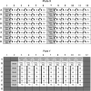
Plate layout for microneutralization assay. Indicated dilutions are suggestions for testing serum samples. Other dilution series or concentrations (for mAbs) might be used.
Figure 3
Figure 3
Analysis of data as reciprocal dilution of serum and percent inhibition of virus (ID50 of serum sample 1 is 1:298 and serum sample 2 is 1:1873).
Figure 4
Figure 4
Plate X and plate Y layout for the antiviral assay.

Similar articles

Cited by

References

    1. Amanat, F. , & Krammer, F. (2020). Sars‐Cov‐2 vaccines: Status report. Immunity, 52(4), 583–589. doi: 10.1016/j.immuni.2020.03.007. - DOI - PMC - PubMed
    1. Amanat, F. , Meade, P. , Strohmeier, S. , & Krammer, F. (2019). Cross‐reactive antibodies binding to H4 hemagglutinin protect against a lethal H4n6 influenza virus challenge in the mouse model. Emerging Microbes & Infections, 8(1), 155–168. doi: 10.1080/22221751.2018.1564369. - DOI - PMC - PubMed
    1. Amanat, F. , Stadlbauer, D. , Strohmeier, S. , Nguyen, T. H. O. , Chromikova, V. , McMahon, M. , … Krammer, F. (2020). A serological assay to detect Sars‐Cov‐2 seroconversion in humans. Nature Medicine. doi: 10.1038/s41591-020-0913-5 [Online ahead of print]. - DOI - PMC - PubMed
    1. Bouhaddou, M. , Memon, D. , Meyer White, K. M. , Rezelj, V. V. , Marrero, M. C. , … Kroger, N. J. The global phosphorylation landscape of SARS‐CoV‐2 infection. Submitted for publication. - PMC - PubMed
    1. Burnett, L. C. , Lunn, G. , & Coico, R. (2009). Biosafety: Guidelines for working with pathogenic and infectious microorganisms. Current Protocols in Microbiology, 13, 1A.1.1–1A.1.14. doi: 10.1002/9780471729259.mc01a01s13. - DOI - PMC - PubMed

Substances

LinkOut - more resources