

Targeting anti-cancer agents to bone using bisphosphonates
- PMID: 32585321
- PMCID: PMC8485333
- DOI: 10.1016/j.bone.2020.115492
Targeting anti-cancer agents to bone using bisphosphonates
Abstract
The skeleton is affected by numerous primary and metastatic solid and hematopoietic malignant tumors, which can cause localized sites of osteolysis or osteosclerosis that can weaken bones and increase the risk of fractures in affected patients. Chemotherapeutic drugs can eliminate some tumors in bones or reduce their volume and skeletal-related events, but adverse effects on non-target organs can significantly limit the amount of drug that can be administered to patients. In these circumstances, it may be impossible to deliver therapeutic drug concentrations to tumor sites in bones. One attractive mechanism to approach this challenge is to conjugate drugs to bisphosphonates, which can target them to bone where they can be released at diseased sites. Multiple attempts have been made to do this since the 1990s with limited degrees of success. Here, we review the results of pre-clinical and clinical studies made to target FDA-approved drugs and other antineoplastic small molecules to bone to treat diseases affecting the skeleton, including osteoporosis, metastatic bone disease, multiple myeloma and osteosarcoma. Results to date are encouraging and indicate that drug efficacy can be increased and side effects reduced using these approaches. Despite these successes, challenges remain: no drugs have gone beyond small phase 2 clinical trials, and major pharmaceutical companies have shown little interest in the approach to repurpose any of their drugs or to embrace the technology. Nevertheless, interest shown by smaller biotechnology companies in the technology suggests that bone-targeting of drugs with bisphosphonates has a viable future.
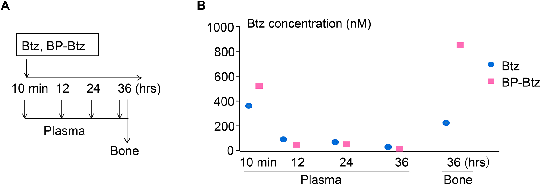
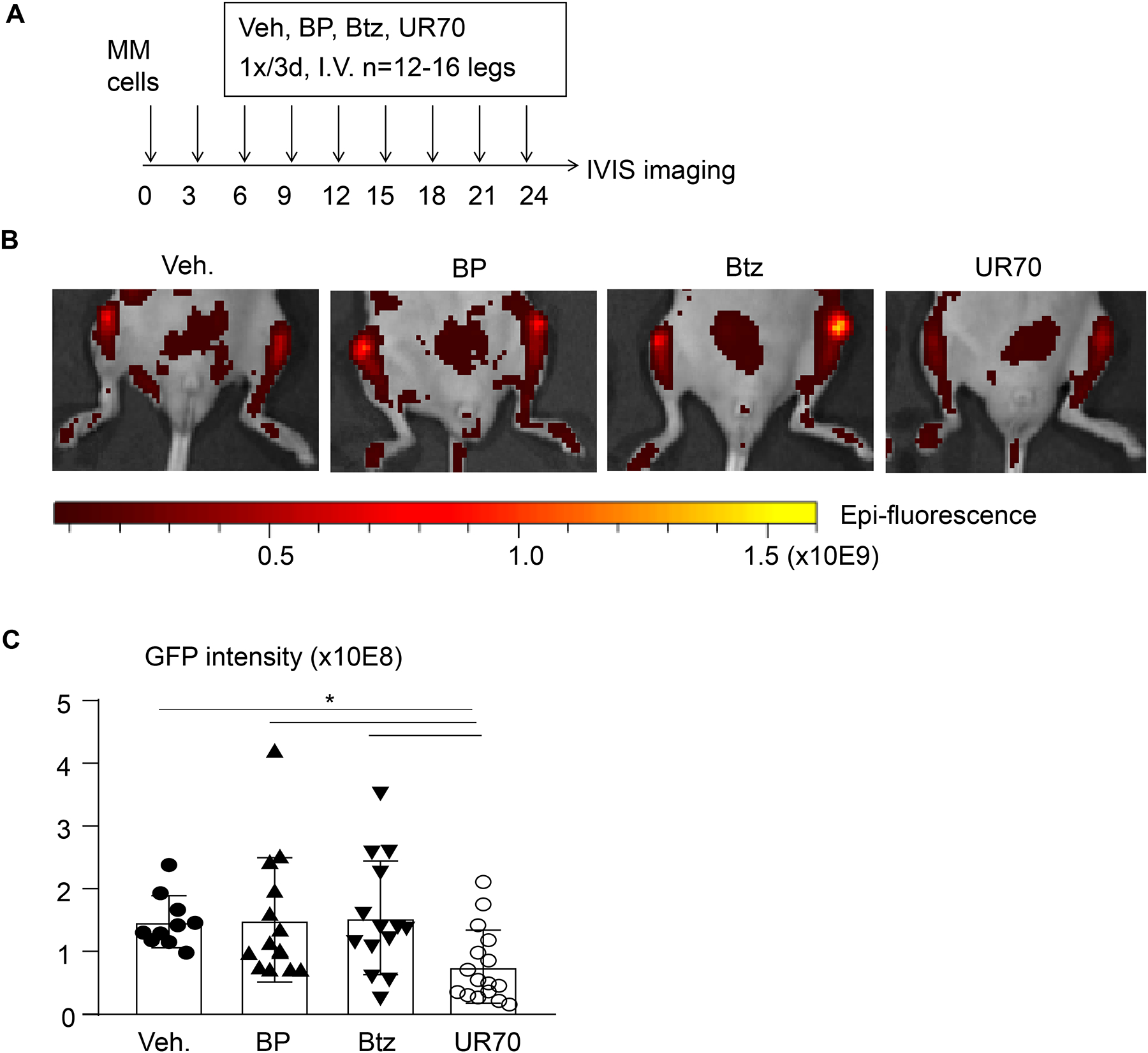
Keywords: Bisphosphonate; Bone metastasis; Bone-targeting; Bortezomib; Chloroquine; Drug conjugate.
Copyright © 2020. Published by Elsevier Inc.
Figures
Similar articles
-
Anti-tumour effects of bisphosphonates--what have we learned from in vivo models?Curr Cancer Drug Targets. 2009 Nov;9(7):807-23. doi: 10.2174/156800909789760339. Curr Cancer Drug Targets. 2009. PMID: 20025569 Review.
-
Mechanisms of action of bisphosphonates on tumor cells and prospects for use in the treatment of malignant osteolysis.Joint Bone Spine. 2000 Jan;67(1):22-9. Joint Bone Spine. 2000. PMID: 10773965 Review.
-
In vitro and in vivo antiresorptive effects of bisphosphonates in metastatic bone disease.In Vivo. 2005 Jan-Feb;19(1):311-8. In Vivo. 2005. PMID: 15796191 Review.
-
[Bone targeting agents: bisphosphonates].Bull Cancer. 2013 Nov;100(11):1199-206. doi: 10.1684/bdc.2013.1840. Bull Cancer. 2013. PMID: 24158668 Review. French.
-
Bisphosphonates in oncology.Bone. 2011 Jul;49(1):71-6. doi: 10.1016/j.bone.2011.02.003. Epub 2011 Feb 21. Bone. 2011. PMID: 21320652 Review.
Cited by
-
Evidence of bisphosphonate-conjugated sitafloxacin eradication of established methicillin-resistant S. aureus infection with osseointegration in murine models of implant-associated osteomyelitis.Bone Res. 2023 Oct 18;11(1):51. doi: 10.1038/s41413-023-00287-4. Bone Res. 2023. PMID: 37848449 Free PMC article.
-
Efficacy of Bisphosphonate-Conjugated Sitafloxacin in a Murine Model of S. aureus Osteomyelitis: Evidence of "Target & Release" Kinetics and Killing of Bacteria Within Canaliculi.Front Cell Infect Microbiol. 2022 Jun 24;12:910970. doi: 10.3389/fcimb.2022.910970. eCollection 2022. Front Cell Infect Microbiol. 2022. PMID: 35811672 Free PMC article.
-
Gemcitabine-Ibandronate Conjugate Enables the Bone-Targeted Combination Therapy in Bone Cancer: Synthesis and Efficacy in Combination with Docetaxel.Bioconjug Chem. 2021 Dec 15;32(12):2530-2539. doi: 10.1021/acs.bioconjchem.1c00507. Epub 2021 Nov 15. Bioconjug Chem. 2021. PMID: 34779607 Free PMC article.
-
Harnessing curcumin and nanotechnology for enhanced treatment of breast cancer bone metastasis.Discov Nano. 2024 Nov 11;19(1):177. doi: 10.1186/s11671-024-04126-1. Discov Nano. 2024. PMID: 39527354 Free PMC article. Review.
-
Hydroxychloroquine and a low antiresorptive activity bisphosphonate conjugate prevent and reverse ovariectomy-induced bone loss in mice through dual antiresorptive and anabolic effects.Bone Res. 2024 Sep 5;12(1):52. doi: 10.1038/s41413-024-00352-6. Bone Res. 2024. PMID: 39231935 Free PMC article.
References
-
- Cetin K, Christiansen CF, Svaerke C, Jacobsen JB, and Sorensen HT, Survival in patients with breast cancer with bone metastasis: a Danish population-based cohort study on the prognostic impact of initial stage of disease at breast cancer diagnosis and length of the bone metastasis-free interval. BMJ Open, 2015. 5(4): p. e007702. DOI: 10.1136/bmjopen-2015-007702. - DOI - PMC - PubMed
Publication types
MeSH terms
Substances
Grants and funding
LinkOut - more resources
Full Text Sources
Other Literature Sources
Medical
