. 2020 Jul 24;23(7):101255.
doi: 10.1016/j.isci.2020.101255. Epub 2020 Jun 10.

The Accumulation of Tau-Immunoreactive Hippocampal Granules and Corpora Amylacea Implicates Reactive Glia in Tau Pathogenesis during Aging

Affiliations

The Accumulation of Tau-Immunoreactive Hippocampal Granules and Corpora Amylacea Implicates Reactive Glia in Tau Pathogenesis during Aging

Connor M Wander et al. iScience. .

Abstract

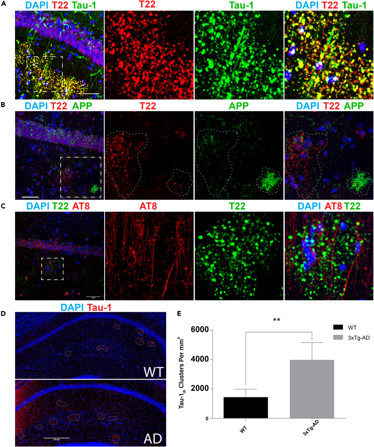
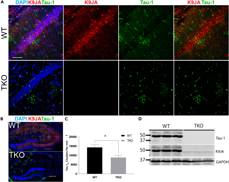
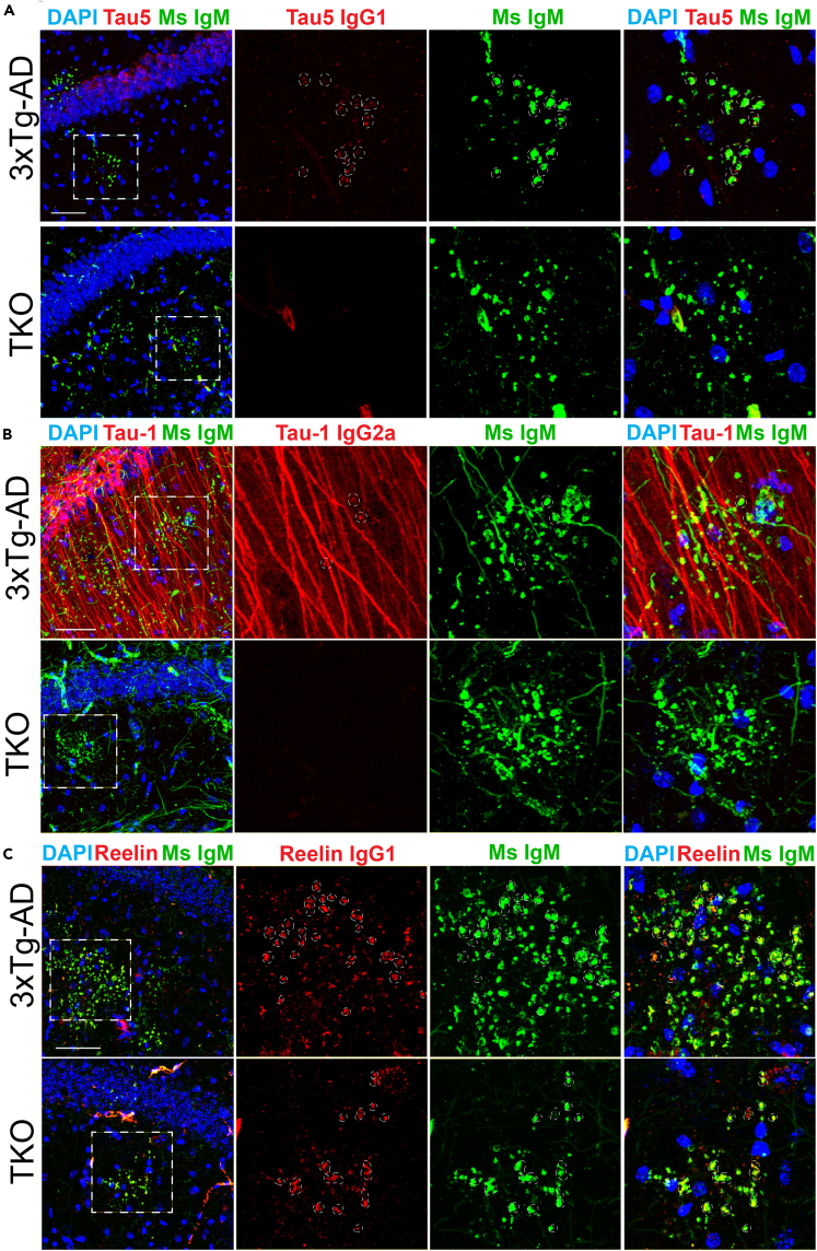
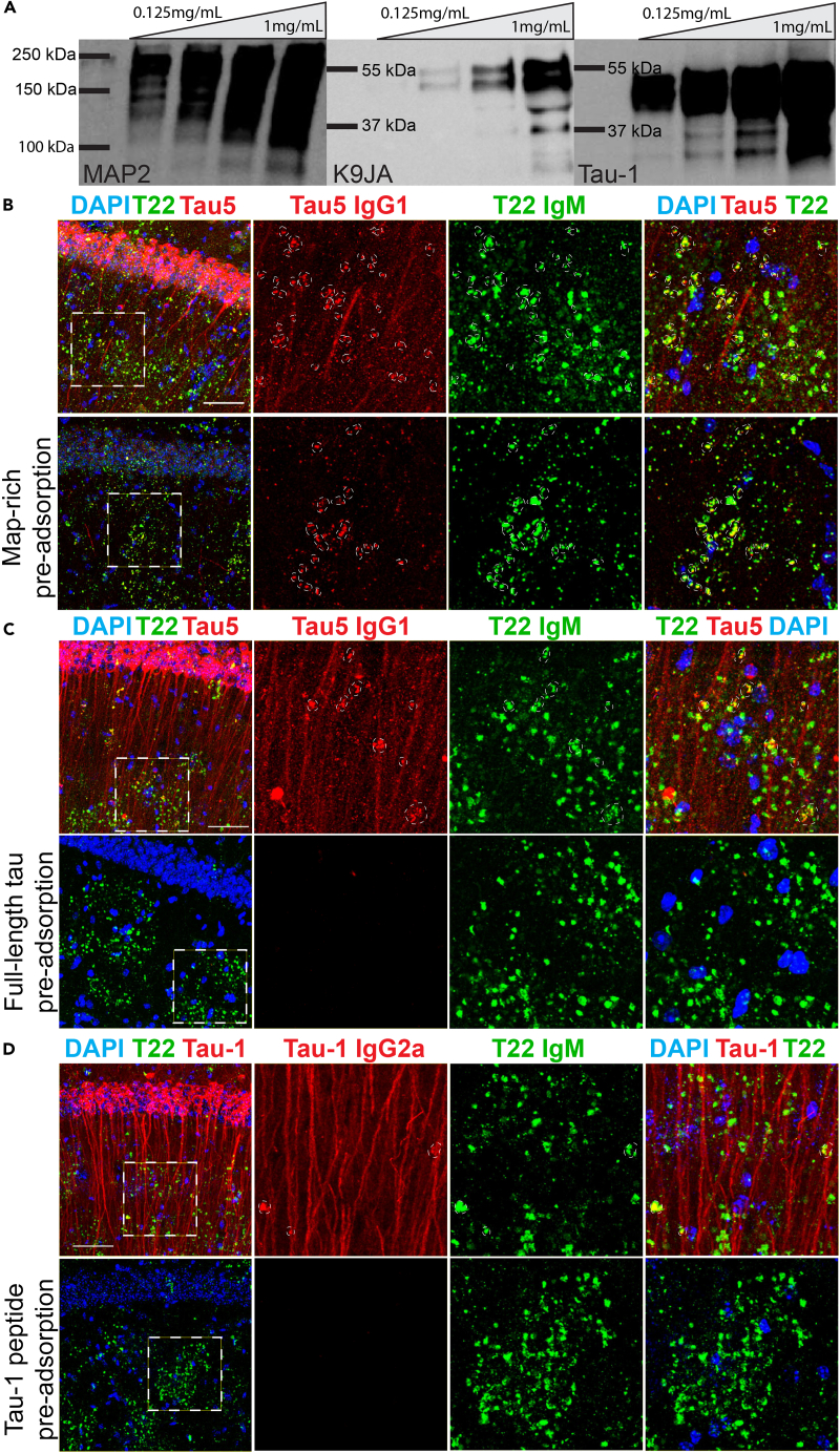
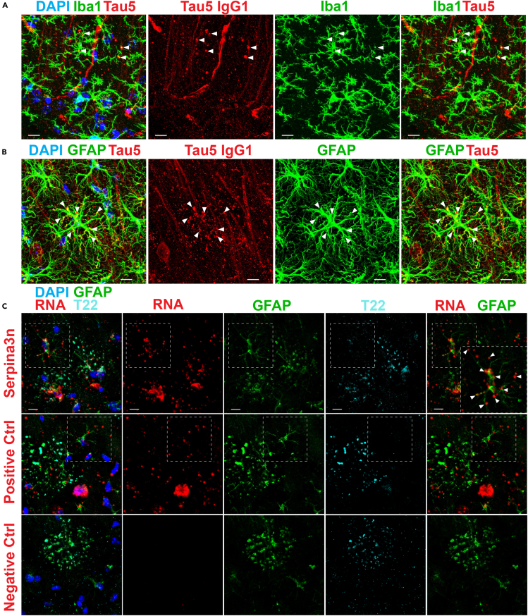
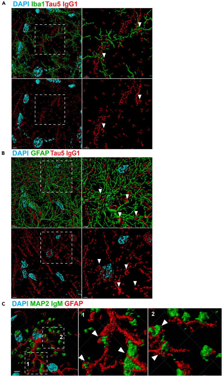
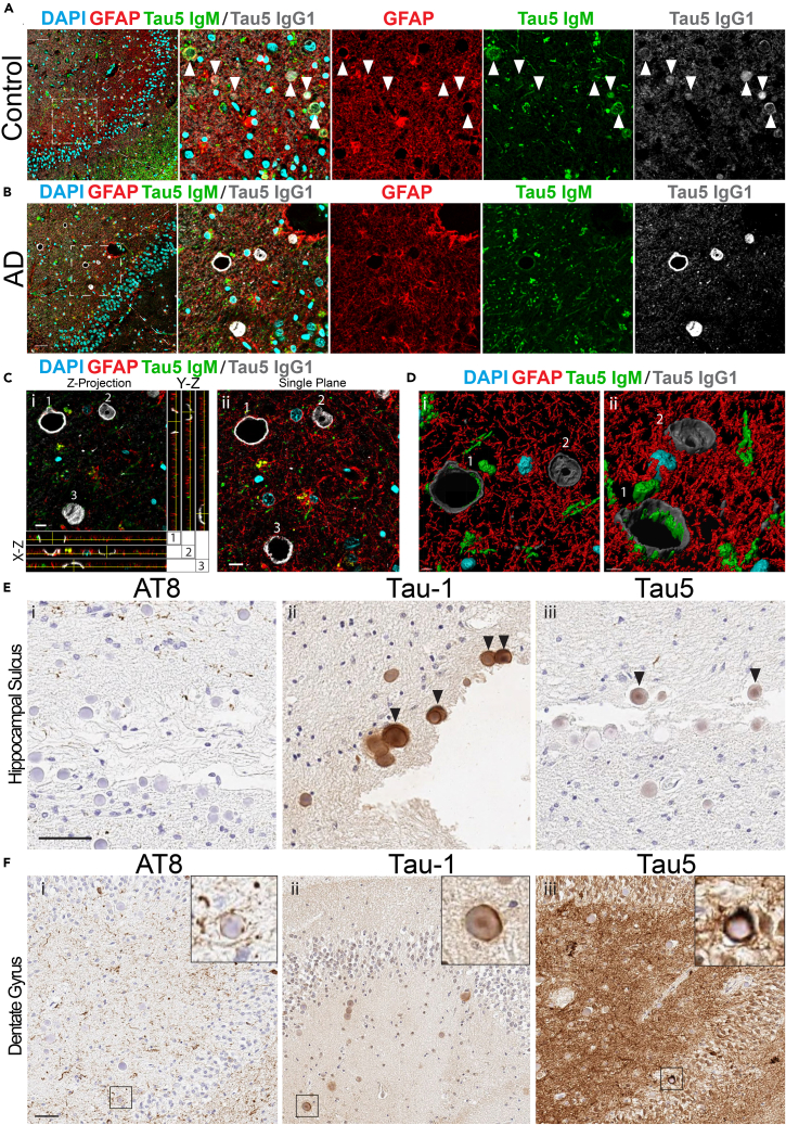
The microtubule-associated tau protein forms pathological inclusions that accumulate in an age-dependent manner in tauopathies including Alzheimer's disease (AD). Since age is the major risk factor for AD, we examined endogenous tau species that evolve during aging in physiological and diseased conditions. In aged mouse brain, we found tau-immunoreactive clusters embedded within structures that are reminiscent of periodic acid-Schiff (PAS) granules. We showed that PAS granules harbor distinct tau species that are more prominent in 3xTg-AD mice. Epitope profiling revealed hypo-phosphorylated rather than hyper-phosphorylated tau commonly observed in tauopathies. High-resolution imaging and 3D reconstruction suggest a link between tau clusters, reactive astrocytes, and microglia, indicating that early tau accumulation may promote neuroinflammation during aging. Using postmortem human brain, we identified tau as a component of corpora amylacea (CA), age-related structures that are functionally analogous to PAS granules. Overall, our study supports neuroimmune dysfunction as a precipitating event in tau pathogenesis.

Keywords: Cellular Neuroscience; Molecular Neuroscience; Neuroscience.


Conflict of interest statement

Declaration of Interests The authors declare no competing interests.

Figures

None
Graphical abstract
Figure 1
Figure 1
Tau-Immunoreactive Clusters in the Hippocampus Are Distinct from Plaque Pathology in 3xTg-AD Mice (A) Immunofluorescent confocal images of aged (21-month-old [mo]) 3xTg-AD mice indicate that Tau-1 (green) and T22 (red) co-localize in granules in the CA1 and SR. (B) T22 (red) does not co-localize with plaques (6E10) or neuronal soma (green) within the CA1 and SR. (C) AT8-immunoreactive neuronal soma and axons (red) associate, but do not co-localize, with T22-positive granules (green) in the CA1 or SR. (D) Representative immunofluorescent images of 3xTg-AD and WT hippocampal fields captured by confocal microscopy at 40x magnification. Dashed circles indicate tau-1IR clusters (red). (E) Quantification of tau-1IR clusters showing that they are significantly elevated in the hippocampi of 6-mo 3xTg-AD mice when compared with controls. Scale bars, 50 μm (A–C), 250 μm (D). Statistical significance was assessed using Student's t test from N = 4 biological replicates (∗∗p < 0.01). Error bars represent SEM. See also Figure S1.
Figure 2
Figure 2
Tau-Immunoreactive Clusters Are Reduced, but Not Eliminated, in Tau KO Mice When Detected with Pan-Reactive Secondary Antibodies (A) Immunofluorescent confocal images of aged (23-mo) wild-type (WT) and tau knockout (TKO) mice show immunoreactivity with K9JA (red) and tau-1 (green) in the CA1 and SR of WT mice, with only tau-1IR cluster immunoreactivity in TKO mice. (B) Representative images of full hippocampal fields in aged WT and TKO mice captured by confocal microscopy at 40x magnification. (C) Quantification of tau-1IR cluster density showing a reduction in granules in 23-mo TKO mice when compared with age-matched WT mice. (D) Immunoblots of tau (tau-1, K9JA) and GAPDH in 23-mo WT and TKO mouse hippocampal lysates confirm the absence of tau expression in TKO mice. Scale bars, 50 μm (A), 250 μm (B). Statistical significance was assessed using a Student's t test from N = 4 biological replicates (∗p < 0.05). Error bars represent SEM. See also Figure S2.
Figure 3
Figure 3
Analysis of Hippocampal Tau Clusters Using Isotype-Specific Immunodetection (A) Immunofluorescent confocal images of aged (23-mo) 3xTg-AD and TKO mice using isotype-specific secondary antibodies. Tau5 detects tau granules when labeled with an anti-mouse IgM μ-chain-specific secondary antibody (Ms IgM, green) in both genotypes and partially detects tau granules when labeled with an anti-mouse IgG1 γ-chain-specific secondary antibody (Tau5 IgG1, red) in 3xTg-AD but not TKO mice. (B) Tau-1 detects tau granules when labeled with an anti-mouse IgM μ-chain-specific secondary antibody (Ms IgM, green) in both genotypes and partially detects tau granules when labeled with an anti-mouse IgG2a γ-chain-specific secondary antibody (Tau-1 IgG2a, red) in 3xTg-AD mice but not TKO mice. (C) Reelin partially detects granules when labeled with both anti-mouse IgM μ-chain-specific (Ms IgM, green) and anti-mouse IgG1 γ-chain-specific (Reelin IgG1, red) secondary antibodies in both 3xTg-AD and TKO mice. Scale bars, 50 μm; dashed white circles indicate regions of co-localization. See also Figure S3.
Figure 4
Figure 4
Analysis of Tau Clusters Following Epitope Neutralization by Immunoadsorption (A) Immunoblots of MAP-rich fractions (MRFs) from porcine brain that were used to pre-adsorb tau-1, tau5, and MAP2 primary antibodies showing MAP2 and tau (K9JA, tau-1) immunoreactivity. (B) The tau5 antibody detects granules when labeled with an anti-mouse IgM μ-chain-specific secondary antibody (Ms IgM, green), labels both dendrites and granules when labeled with an anti-mouse IgG1-specific secondary antibody (Tau5 IgG1, red), and is partially pre-adsorbed when neutralized with MRF, whereas anti-mouse IgM-specific granules (Ms IgM, green) remain in 21-mo 3xTg-AD mice. (C) Tau5 anti-mouse IgG1-specific (Tau5 IgG1, red) immunoreactivity is neutralized with recombinant full-length (2N4R isoform) tau protein, whereas anti-mouse IgM-specific granules (green) remain in 21-mo 3xTg-AD mice. (D) The tau-1 antibody detects granules when labeled with anti-mouse IgM μ-chain-specific secondary antibody (Ms IgM, green), detects dendrites and some, but not all, granules when labeled with anti-IgG2a secondary antibody (Tau-1 IgG2a, red) in 21-mo 3xTg-AD mice. Tau-1 anti-mouse IgG2a-specific immunoreactivity is neutralized by the purified tau-1 peptide, whereas anti-mouse IgM-specific (Ms IgM, green) immunoreactivity remains. Scale bars, 50 μm, dashed white circles indicate regions of co-localization. See also Figure S4.
Figure 5
Figure 5
Tau-Immunoreactive Granules Are Associated with Reactive Astrocytes (A) Immunofluorescent confocal images of aged (21-mo) 3xTg-AD mice show distal processes of microglia detected with anti-rabbit Iba1 (Iba1, green) that associate with and envelop anti-mouse IgG1-specific tau5IR granules (Tau5 IgG1, red) in the CA1 and SR of aged 3xTg-AD mice. (B) Distal processes of astrocytes detected with anti-rabbit GFAP (GFAP, green) contain and envelop many anti-mouse IgG1-specific tau5IR granules (Tau5 IgG1, red) in the CA1 and SR of aged 3xTg-AD mice. (C) Confocal images of dual in situ RNA labeling and immunofluorescence shows Serpina3n expression (red) in processes of reactive astrocytes (GFAP, green) entangled with T22-positive hippocampal clusters (T22, cyan). Robust expression of Ppib (positive control, red) is also detected in affected astrocytes (green) and other cells (DAPI, blue), whereas the negative control probe (red) is not detected in the hippocampus. Scale bars, 10 μm; arrowheads indicate sites of co-localization. See also Figure S5.
Figure 6
Figure 6
3D Reconstruction of Tau-Immunoreactive Granules Reveals Extensive Association with Microglia and Astrocytes (A) 3D reconstruction of confocal images obtained at 60x magnification (with 2× zoom) show that Iba1-positive microglial processes (green) associate with anti-mouse IgG-specific tau5IR dendrites and granules (Tau5 IgG1, red) in the SR of 3xTg-AD mice. (B) 3D reconstruction shows that GFAP-positive astrocytic processes (green) associated with anti-mouse IgG-specific tau5IR dendrites and granules (Tau5 IgG1, red) in the SR of 3xTg-AD mice. (C) 3D reconstruction shows that anti-mouse IgM-specific granules from the mouse monoclonal MAP2 primary antibody (MAP2 IgM, green) are closely juxtaposed along processes of GFAP-positive astrocytes (red). Scale bar, 5 μm (A, B, left); 3 μm (A, B, right); 5 μm (C, left). Arrowheads indicate regions of association.
Figure 7
Figure 7
Tau-Immunoreactive CA in Human Control and AD Brain (A) Confocal images of human control hippocampus immunostained with tau5 and GFAP primary antibodies and anti-mouse IgM μ-chain (Tau5 IgM, green), anti-rabbit (GFAP, red), and anti-mouse IgG1 γ-chain-specific (Tau5 IgG1, gray) secondary antibodies. Tau5 detects tau within CA when labeled with an anti-mouse IgG1 secondary antibody (gray). CA were labeled with an anti-mouse IgM μ-chain-specific secondary antibody (green). GFAP surrounds CA when labeled with anti-rabbit secondary antibody (red). Scale bar, 50 μm (arrowheads indicate immunoreactive CA). (B) Confocal images of human AD hippocampus immunostained with tau5 and GFAP primary antibodies and isotype-specific secondaries detailed above. Tau5 detects tau within CA when labeled with an anti-mouse IgG1 secondary antibody (gray). CA were labeled with an anti-mouse IgM-specific secondary antibody (green). GFAP outlines but does not fill CA when labeled with an anti-rabbit secondary antibody (red). Scale bar, 50 μm. (C) Average-intensity Z projection from selected region in (B), surrounded by orthogonal views targeted to individual CA labeled as 1, 2, and 3 (i). A single plane confocal image of AD patient hippocampus is also shown (ii). Scale bar, 10 μm. (D) 3D reconstruction of inset showing hollowed structure of CA from (i) top-down and (ii) oblique angular view. (E) Wide-field IHC images of the hippocampal sulcus in serial sections from an AD patient brain stained with (i) AT8, (ii) Tau-1, and (iii) Tau5 primary antibodies and anti-mouse IgG1-specific or anti-mouse IgG2a-specific HRP-conjugated secondary antibodies. Scale bar, 50 μm. Arrowheads indicate tau-immunoreactive CA. (F) Wide-field IHC images of the dentate gyrus in serial sections from AD patient brain stained with (i) AT8, (ii) tau-1, and (iii) tau5 primary antibodies and detected with anti-mouse IgG1-specific or anti-mouse IgG2a-specific HRP-conjugated secondary antibodies. Scale bar, 50 μm. See also Figures S6 and S7 and Table S3.

References

    1. Akiyama H., Kameyama M., Akiguchi I., Sugiyama H., Kawamata T., Fukuyama H., Kimura H., Matsushita M., Takeda T. Periodic acid-Schiff (PAS)-positive, granular structures increase in the brain of senescence accelerated mouse (SAM) Acta Neuropathol. 1986;72:124–129. - PubMed
    1. Auge E., Bechmann I., Llor N., Vilaplana J., Krueger M., Pelegri C. Corpora amylacea in human hippocampal brain tissue are intracellular bodies that exhibit a homogeneous distribution of neo-epitopes. Sci. Rep. 2019;9:2063. - PMC - PubMed
    1. Auge E., Cabezon I., Pelegri C., Vilaplana J. New perspectives on corpora amylacea in the human brain. Sci. Rep. 2017;7:41807. - PMC - PubMed
    1. Auge E., Duran J., Guinovart J.J., Pelegri C., Vilaplana J. Exploring the elusive composition of corpora amylacea of human brain. Sci. Rep. 2018;8:13525. - PMC - PubMed
    1. Baker C., Belbin O., Kalsheker N., Morgan K. SERPINA3 (aka alpha-1-antichymotrypsin) Front. Biosci. 2007;12:2821–2835. - PubMed

LinkOut - more resources