. 2020 Jun 23;25(12):2882.
doi: 10.3390/molecules25122882.

Gas Sensing by Bacterial H-NOX Proteins: An MD Study

Affiliations

Gas Sensing by Bacterial H-NOX Proteins: An MD Study

Ahmed M Rozza et al. Molecules. .

Abstract

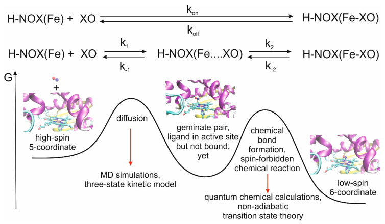
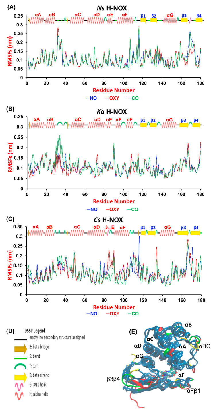
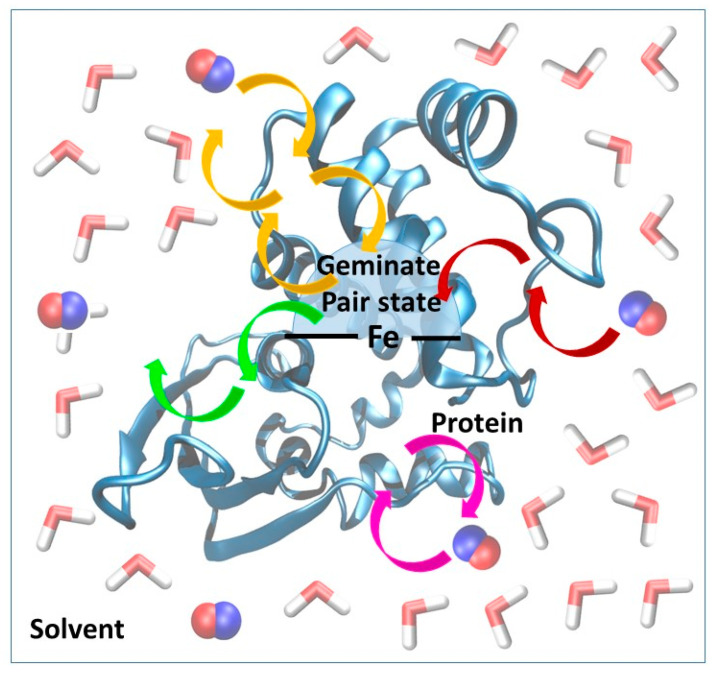
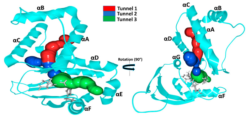
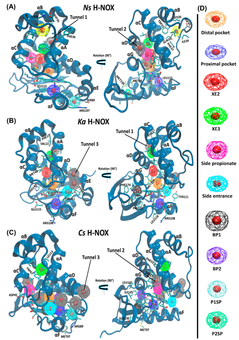
Gas sensing is crucial for both prokaryotes and eukaryotes and is primarily performed by heme-based sensors, including H-NOX domains. These systems may provide a new, alternative mode for transporting gaseous molecules in higher organisms, but for the development of such systems, a detailed understanding of the ligand-binding properties is required. Here, we focused on ligand migration within the protein matrix: we performed molecular dynamics simulations on three bacterial (Ka, Ns and Cs) H-NOX proteins and studied the kinetics of CO, NO and O2 diffusion. We compared the response of the protein structure to the presence of ligands, diffusion rate constants, tunnel systems and storage pockets. We found that the rate constant for diffusion decreases in the O2 > NO > CO order in all proteins, and in the Ns > Ks > Cs order if single-gas is considered. Competition between gases seems to seriously influence the residential time of ligands spent in the distal pocket. The channel system is profoundly determined by the overall fold, but the sidechain pattern has a significant role in blocking certain channels by hydrophobic interactions between bulky groups, cation-π interactions or hydrogen bonding triads. The majority of storage pockets are determined by local sidechain composition, although certain functional cavities, such as the distal and proximal pockets are found in all systems. A major guideline for the design of gas transport systems is the need to chemically bind the gas molecule to the protein, possibly joining several proteins with several heme groups together.

Keywords: H-NOX; diffusion; gas sensing; heme protein; migration routes; molecular dynamics; sGC.


Conflict of interest statement

The authors declare no conflicts of interest.

Figures

Figure 1
Figure 1
Ligand binding by Heme-Nitric oxide and OXygen (H-NOX) proteins and definition of the relevant rate constants. Experimentally, and usually, kon and koff are determined, while calculations allow us to study the steps individually and obtain the values of k1, k−1, k2 and k−2. The overall process can be divided into two major events: diffusion of the ligand to the active site and the formation of the bond between the ligand and the Fe(II) ion. Due to the nature of the two events, different methodologies are required for their proper description, and as the spins are inverted on iron in the course of the chemical reaction, a special form of transition state theory is needed for the derivation of the rate constant values [15,22].
Figure 2
Figure 2
RMSF values computed per-residue from molecular dynamics (MD) simulations of gaseous molecule (NO, CO, O2) diffusion into (A) Nostoc sp. (Ns), (B) Kordia algicida (Ka) and (C) Caldanaerobacter subterraneus (Cs) H-NOX. In the case of Cs, the RMSF values of the long tail (residues 181–188 that are not present in Ns and Ka H-NOX) are not shown to make comparison with other systems easier. (D) Labels used for the designation of secondary structure elements by DSPP in Figure 2A–C. (E) Superposition of the structure of the three H-NOX proteins. Regions with highest mobility have been highlighted by larger font size, and colored in green (Ns), yellow (Ka) and red (Cs).
Figure 3
Figure 3
Schematic representation of the kinetic model used for the estimation of the rate constants for diffusion. The overall system has been divided into three parts: solvent phase (water molecules are shown in licorice), protein and geminate pair state (the geminate pair state is slightly smaller than the distal pocket). The heme group is represented by solid black lines. Ligand molecules (shown in van der Waals representation) may enter and leave the protein via various routes (indicated by yellow, green, pink and red arrows.). In the model, only those events are counted which represent the migration of the ligand from the solvent phase via the protein to the distal pocket and back to the solvent. Oscillations between the protein and the solvent/distal pocket are not taken into account.
Figure 4
Figure 4
Identified gas migration tunnels. As the three-dimensional structures of the three proteins are very similar only the structure of Ns H-NOX is shown. The long, apolar tunnel is shown in red, while the shorter Tunnels 2 and 3 are shown in blue and green, respectively. The heme group and the proximal histidine residue are shown in gray.
Figure 5
Figure 5
Gas-binding cavities in the studied H-NOX systems. Color coding used for the cavities are shown in the panel on the right.

References

    1. Tsai A.-L., Berka V., Martin F., Ma X., van den Akker F., Fabian M., Olson J.S. Is Nostoc H-NOX an NO sensor or Redox Switch? Biochemistry. 2010;49:6587–6599. doi: 10.1021/bi1002234. - DOI - PMC - PubMed
    1. Liao M.-S., Huang M.-J., Watts J.D. Binding of O2 and NO to Heme in Heme-Nitric Oxide/OXygen- Binding (H-NOX) Proteins. A Theoretical Study. J. Phys. Chem. B. 2013;117:10103–10114. doi: 10.1021/jp403998u. - DOI - PMC - PubMed
    1. Kajimura M., Nakanishi T., Takenouchi T., Morikawa T., Hishiki T., Yukutake Y., Suematsu M. Gas biology: Tiny molecules controlling metabolic systems. Respir. Physiol. Neurobiol. 2012;184:139–148. doi: 10.1016/j.resp.2012.03.016. - DOI - PubMed
    1. Pan J., Yuan H., Zhang X., Zhang H., Xu Q., Zhou Y., Tan L., Nagawa S., Huang Z.X., Tan X. Probing the Molecular Mechanism of Human Soluble Guanylate Cyclase Activation by NO in vitro and in vivo. Sci. Rep. 2017;7:1–14. doi: 10.1038/srep43112. - DOI - PMC - PubMed
    1. Mustafa A.K., Gadalla M.M., Snyder S.H. Signaling by Gasotransmitters. Bone. 2009;23:1–7. doi: 10.1126/scisignal.268re2. - DOI - PMC - PubMed