Review
. 2020 Jun 23;25(12):2881.
doi: 10.3390/molecules25122881.

Structural Insights into RNA Dimerization: Motifs, Interfaces and Functions

Affiliations
Review

Structural Insights into RNA Dimerization: Motifs, Interfaces and Functions

Charles Bou-Nader et al. Molecules. .

Abstract

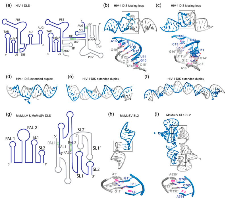
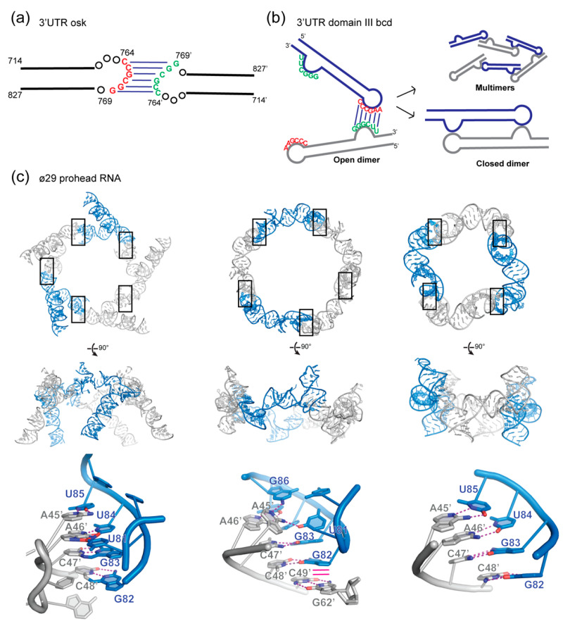
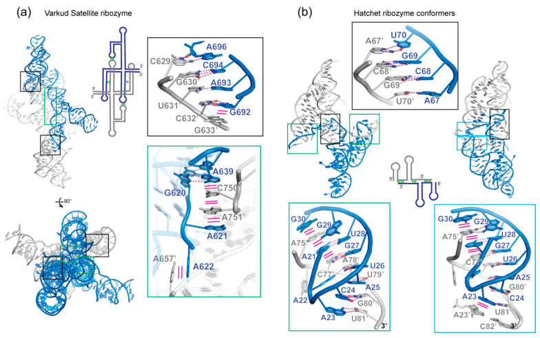
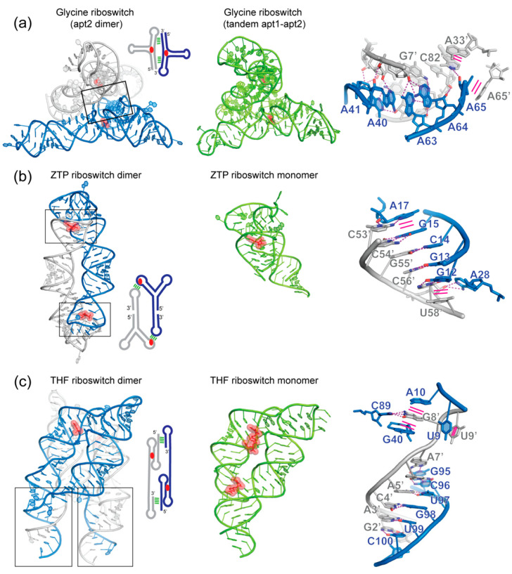
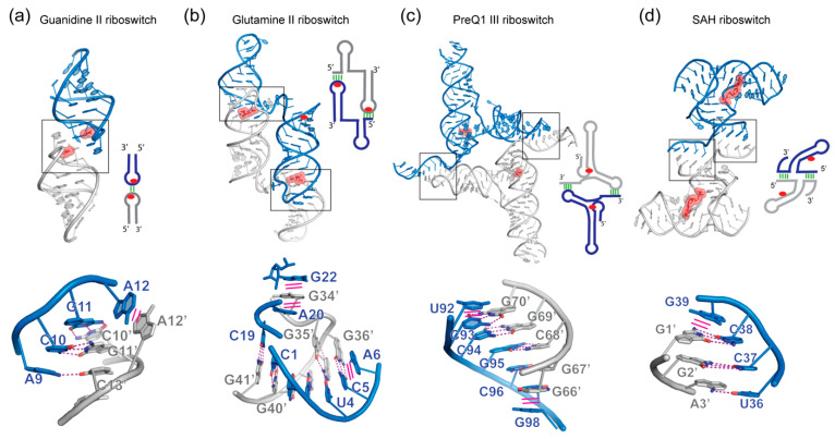
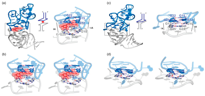
In comparison with the pervasive use of protein dimers and multimers in all domains of life, functional RNA oligomers have so far rarely been observed in nature. Their diminished occurrence contrasts starkly with the robust intrinsic potential of RNA to multimerize through long-range base-pairing ("kissing") interactions, self-annealing of palindromic or complementary sequences, and stable tertiary contact motifs, such as the GNRA tetraloop-receptors. To explore the general mechanics of RNA dimerization, we performed a meta-analysis of a collection of exemplary RNA homodimer structures consisting of viral genomic elements, ribozymes, riboswitches, etc., encompassing both functional and fortuitous dimers. Globally, we found that domain-swapped dimers and antiparallel, head-to-tail arrangements are predominant architectural themes. Locally, we observed that the same structural motifs, interfaces and forces that enable tertiary RNA folding also drive their higher-order assemblies. These feature prominently long-range kissing loops, pseudoknots, reciprocal base intercalations and A-minor interactions. We postulate that the scarcity of functional RNA multimers and limited diversity in multimerization motifs may reflect evolutionary constraints imposed by host antiviral immune surveillance and stress sensing. A deepening mechanistic understanding of RNA multimerization is expected to facilitate investigations into RNA and RNP assemblies, condensates, and granules and enable their potential therapeutical targeting.

Keywords: RNA; dimerization; domain swapping; folding; intermolecular interaction; riboswitches; ribozymes; structure.


Conflict of interest statement

The authors declare no conflict of interest.

Figures

Figure 1
Figure 1
Homodimerization of retroviral genomic RNAs (gRNA). (a) Schematic view of the 5′ leader dimer linkage site (DLS) of human immunodeficiency virus type 1 (HIV-1) driving gRNA dimerization. Left: monomeric state with the dimerization initiation site (DIS) sequestrated by the U5 segment. Right: dimeric state driven by intermolecular DIS-DIS’ interaction. The Gag start site AUG’ element pairs in trans with the U5 segment. These conformational changes allow the HIV-1 genome to switch between translation (monomeric) and packaging (dimeric) functions. Structural elements followed by a prime symbol indicate that they belong to the other protomer. (b,c) Crystal and NMR structure of the DIS kissing loop interaction, respectively (PDB 2B8S and 1BAU). (d,e,f) Crystal, NMR, and Cryo-EM structures of the DIS homodimer in its extended duplex state (PDB 1Y99, 2GM0 and 6BG9). (g) Schematic view of the 5′ leader structure of the Moloney murine leukemia virus (MoMuLV) and Moloney murine sarcoma virus (MoMuSV) regulating gRNA dimerization. Four elements in this RNA drive its dimerization: palindromic sequences in PAL1 and PAL2 and kissing loop interactions between Stem-loop 1 (SL1) and Stem-loop 2 (SL2). (h) Structure of MoMuLV homodimer SL2 (PDB 1F5U). (i) Structure of MoMuSV homodimer SL1-SL2 (PDB 2L1F). Interface regions are boxed and detailed below each structure. Hydrogen bonding interactions are shown as magenta dashed lines, while stacking interactions are shown as solid magenta lines.
Figure 2
Figure 2
Homodimerization in mRNA transport. (a) oskar mRNA dimerize through a conserved GC rich kissing loop in the 3′-untranslated region (UTR). (b) bicoid mRNA dimerization motif relies on the complementarity of the apical loop and an internal bulge in the domain III of its 3′-UTR. This leads to formation of closed dimers or open dimers that can further oligomerize. (c) Structures of the prohead RNA of bacteriophage ø29 driven by kissing interactions between two stem-loops. From left to right, two cryo-EM structures of the pentameric assembly of pRNA (PDB 1FOQ and 6QYZ) and a crystal structure of a pRNA tetramer (PDB 3R4F). The conserved feature amongst the three structures is the Watson-Crick pairing between the 45AACC48 and 82GGUU85 segments, which drives higher-order assembly. Interface regions are boxed and detailed below each structure. Hydrogen bonding interactions are shown as magenta dashed lines, while stacking interactions are shown as solid magenta lines.
Figure 3
Figure 3
Structures of dimeric ribozymes. (a) Crystal structure of Varkud Satellite ribozyme (PDB code 4R4P & 4R4V) with the insets showing the intermolecular kissing loop interaction between stem-loop 1 (P1) and stem-loop 5′ (P5′), as well as the intercalation of stacked nucleobases between P1 and P6′. (b) Crystal structures of the Hatchet ribozyme (PDB 6QJ6 & 6QJ5) showing two types of dimeric interfaces. The top inset highlights the palindromic sequence paired with the same sequence of the second protomer. Hydrogen bonding interactions are shown as magenta dashed lines, while stacking interactions are shown as solid magenta lines.
Figure 4
Figure 4
Comparison of monomeric and dimeric structures of the same riboswitches. (a) Glycine aptamer 2 crystallized in a dimer (PDB 3OWI) configuration similar to the tandem glycine aptamer1-aptamer2 (PDB 3P49). (b) ZTP riboswitch dimer structure (PDB 4XWF) juxtaposed with its monomeric version (PDB 5BTP). (c) THF riboswitch crystallized in dimeric (PDB 3SUY and 3SUX) and monomeric version (PDB 4LVV). Ligands are shown in stick representation and semi-transparent red spheres. Interface regions are boxed and detailed on the right of each dimeric structure. Hydrogen bonding interactions are shown as magenta dashed lines, and stacking interactions are shown as solid magenta lines.
Figure 5
Figure 5
Additional in crystallo dimeric riboswitches. (a) Guanidine II riboswitch (PDB 5NDI and 5VJ9). (b) Glutamine riboswitch II (PDB 6QN3). (c) Class III preQ1 riboswitch (PDB 4RZD). (d) SAH riboswitch (PDB 3NPQ). Ligands are shown in stick representation and semi-transparent red spheres. Interface regions are boxed and detailed below each structure. Hydrogen bonding interactions are shown as magenta dashed lines, and stacking interactions are shown as solid magenta lines.
Figure 6
Figure 6
Dimeric structure of fluorogenic Corn RNA. (a) Bound to the DHFO (3,5-difluoro-4-hydroxybenzylidene- imidazolinone-2-oxime) ligand (PDB 5BJO) with a stereo view of its dimeric interface in (b). (c) In its apo form (PDB 6E80) with a stereo view of the collapsed dimeric interface in the absence of ligand (d). Ligands are shown in stick representation and semi-transparent red spheres. Interface regions are boxed and detailed below each structure. Hydrogen bonding interactions are shown as magenta dashed lines, and stacking interactions are shown as solid magenta lines.

Similar articles

Cited by

References

    1. Levy E.D., Erba B.E., Robinson V.C., Teichmann A.S. Assembly reflects evolution of protein complexes. Nature. 2008;453:1262–1265. doi: 10.1038/nature06942. - DOI - PMC - PubMed
    1. Jones C.P., Ferré-D’Amaré A.R. RNA quaternary structure and global symmetry. Trends Biochem. Sci. 2015;40:211–220. doi: 10.1016/j.tibs.2015.02.004. - DOI - PMC - PubMed
    1. Perica T., Marsh A.J., Sousa L.F., Natan E., Colwell J.L., Ahnert E.S., Teichmann A.S. The emergence of protein complexes: Quaternary structure, dynamics and allostery. Colworth Medal Lecture. Biochem. Soc. Trans. 2012;40:475–491. doi: 10.1042/BST20120056. - DOI - PubMed
    1. Goodsell D.S., Olson A.J. Structural symmetry and protein function. Annu. Rev. Biophys. Biomol. Struct. 2000;29:105–153. doi: 10.1146/annurev.biophys.29.1.105. - DOI - PubMed
    1. Marianayagam N.J., Sunde M., Matthews J.M. The power of two: Protein dimerization in biology. Trends Biochem. Sci. 2004;29:618–625. doi: 10.1016/j.tibs.2004.09.006. - DOI - PubMed

LinkOut - more resources