

Tooth Enamel and its Dynamic Protein Matrix
- PMID: 32585904
- PMCID: PMC7352428
- DOI: 10.3390/ijms21124458
Tooth Enamel and its Dynamic Protein Matrix
Abstract
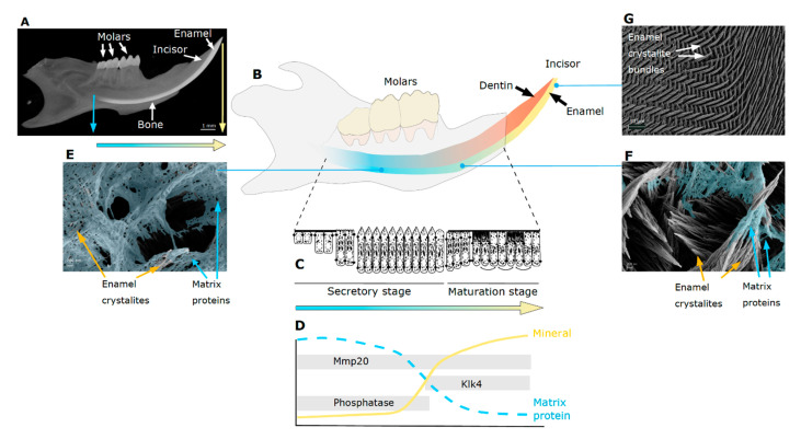
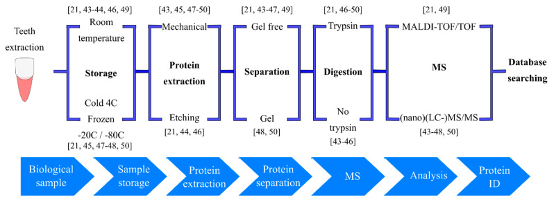
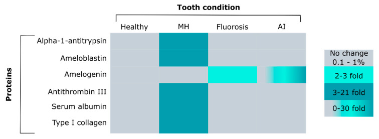
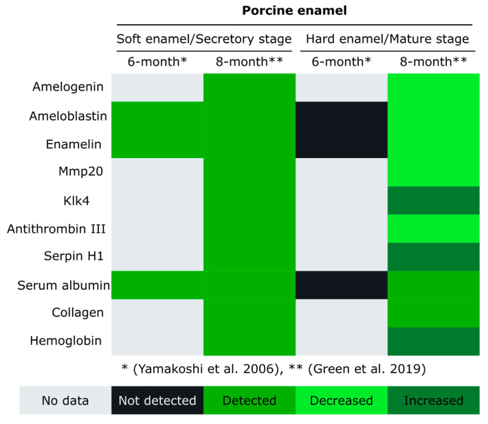
Tooth enamel is the outer covering of tooth crowns, the hardest material in the mammalian body, yet fracture resistant. The extremely high content of 95 wt% calcium phosphate in healthy adult teeth is achieved through mineralization of a proteinaceous matrix that changes in abundance and composition. Enamel-specific proteins and proteases are known to be critical for proper enamel formation. Recent proteomics analyses revealed many other proteins with their roles in enamel formation yet to be unraveled. Although the exact protein composition of healthy tooth enamel is still unknown, it is apparent that compromised enamel deviates in amount and composition of its organic material. Why these differences affect both the mineralization process before tooth eruption and the properties of erupted teeth will become apparent as proteomics protocols are adjusted to the variability between species, tooth size, sample size and ephemeral organic content of forming teeth. This review summarizes the current knowledge and published proteomics data of healthy and diseased tooth enamel, including advancements in forensic applications and disease models in animals. A summary and discussion of the status quo highlights how recent proteomics findings advance our understating of the complexity and temporal changes of extracellular matrix composition during tooth enamel formation.
Keywords: amelogenin; amelogenin-Y (AMELY); dental anthropology; dental fluorosis; enamel peptide; enamel proteome; molar hypomineralization; serum albumin; tooth enamel.
Conflict of interest statement
The authors declare no conflict of interest.
Figures
Similar articles
-
Mapping the Tooth Enamel Proteome and Amelogenin Phosphorylation Onto Mineralizing Porcine Tooth Crowns.Front Physiol. 2019 Jul 30;10:925. doi: 10.3389/fphys.2019.00925. eCollection 2019. Front Physiol. 2019. PMID: 31417410 Free PMC article.
-
Dental fluorosis: chemistry and biology.Crit Rev Oral Biol Med. 2002;13(2):155-70. doi: 10.1177/154411130201300206. Crit Rev Oral Biol Med. 2002. PMID: 12097358 Review.
-
Posteruptive Loss of Proteins in Porcine Enamel.J Dent Res. 2025 Mar;104(3):290-298. doi: 10.1177/00220345241299382. Epub 2024 Dec 26. J Dent Res. 2025. PMID: 39725879
-
Identification of proteins from human permanent erupted enamel.Eur J Oral Sci. 2015 Dec;123(6):390-5. doi: 10.1111/eos.12214. Epub 2015 Oct 3. Eur J Oral Sci. 2015. PMID: 26432388
-
DENTAL ENAMEL FORMATION AND IMPLICATIONS FOR ORAL HEALTH AND DISEASE.Physiol Rev. 2017 Jul 1;97(3):939-993. doi: 10.1152/physrev.00030.2016. Physiol Rev. 2017. PMID: 28468833 Free PMC article. Review.
Cited by
-
Advances in experimental and computational methodologies for the study of microbial-surface interactions at different omics levels.Front Microbiol. 2022 Nov 28;13:1006946. doi: 10.3389/fmicb.2022.1006946. eCollection 2022. Front Microbiol. 2022. PMID: 36519168 Free PMC article. Review.
-
Differential Expression of Hard Tissue Proteins in Hypomineralized Second Primary Molars in Comparison to Normal Teeth.Clin Exp Dent Res. 2025 Feb;11(1):e70079. doi: 10.1002/cre2.70079. Clin Exp Dent Res. 2025. PMID: 39898784 Free PMC article.
-
Effect of erosive conditions on different sealant materials used in paediatric dentistry.Braz Oral Res. 2024 Jun 24;38:e053. doi: 10.1590/1807-3107bor-2024.vol38.0053. eCollection 2024. Braz Oral Res. 2024. PMID: 38922213 Free PMC article.
-
Research progress of biomimetic materials in oral medicine.J Biol Eng. 2023 Nov 23;17(1):72. doi: 10.1186/s13036-023-00382-4. J Biol Eng. 2023. PMID: 37996886 Free PMC article. Review.
-
The Effects of Three Remineralizing Agents on the Microhardness and Chemical Composition of Demineralized Enamel.Materials (Basel). 2021 Oct 13;14(20):6051. doi: 10.3390/ma14206051. Materials (Basel). 2021. PMID: 34683643 Free PMC article.
References
-
- Davis K.A., Mountain R.V., Pickett O.R., Den Besten P.K., Bidlack F.B., Dunn E.C. Teeth as Potential New Tools to Measure Early-Life Adversity and Subsequent Mental Health Risk: An Interdisciplinary Review and Conceptual Model. Biol. Psychiatr. 2020;87:502–513. doi: 10.1016/j.biopsych.2019.09.030. - DOI - PMC - PubMed
-
- Queen Mary University of London—Institute of Dentistry—Barts and The London Atlas of Tooth Development and Eruption. [(accessed on 22 June 2020)]; Available online: https://www.qmul.ac.uk/dentistry/atlas/
-
- Thesleff I. Developmental biology and building a tooth. Quintessence Int. 2003;34:613–620. - PubMed
Publication types
MeSH terms
Substances
Grants and funding
LinkOut - more resources
Full Text Sources
