Review
. 2020 Jun 23;12(6):1668.
doi: 10.3390/cancers12061668.

Glucose Metabolism and Oxidative Stress in Hepatocellular Carcinoma: Role and Possible Implications in Novel Therapeutic Strategies

Affiliations
Review

Glucose Metabolism and Oxidative Stress in Hepatocellular Carcinoma: Role and Possible Implications in Novel Therapeutic Strategies

Monica Mossenta et al. Cancers (Basel). .

Abstract

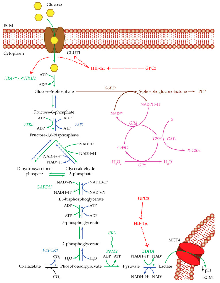
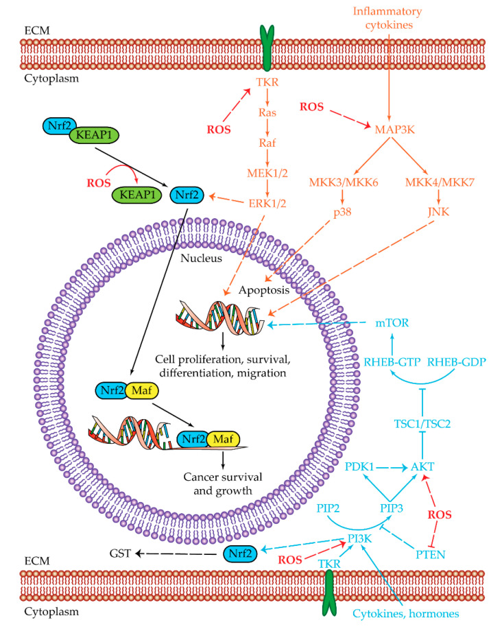
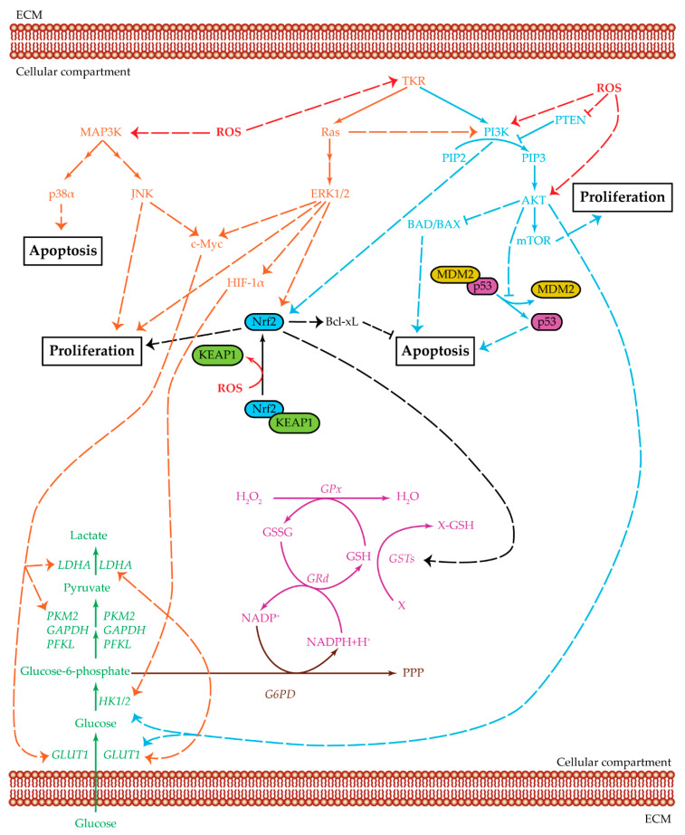
Hepatocellular carcinoma (HCC) metabolism is redirected to glycolysis to enhance the production of metabolic compounds employed by cancer cells to produce proteins, lipids, and nucleotides in order to maintain a high proliferative rate. This mechanism drives towards uncontrolled growth and causes a further increase in reactive oxygen species (ROS), which could lead to cell death. HCC overcomes the problem generated by ROS increase by increasing the antioxidant machinery, in which key mechanisms involve glutathione, nuclear factor erythroid 2-related factor 2 (Nrf2), and hypoxia-inducible transcription factor (HIF-1α). These mechanisms could represent optimal targets for innovative therapies. The tumor microenvironment (TME) exerts a key role in HCC pathogenesis and progression. Various metabolic machineries modulate the activity of immune cells in the TME. The deregulated metabolic activity of tumor cells could impair antitumor response. Lactic acid-lactate, derived from the anaerobic glycolytic rate of tumor cells, as well as adenosine, derived from the catabolism of ATP, have an immunosuppressive activity. Metabolic reprogramming of the TME via targeted therapies could enhance the treatment efficacy of anti-cancer immunotherapy. This review describes the metabolic pathways mainly involved in the HCC pathogenesis and progression. The potential targets for HCC treatment involved in these pathways are also discussed.

Keywords: HCC; anticancer-immunoresponse; glucose metabolism; oxidative stress; tumor microenvironment.


Conflict of interest statement

The authors declare no conflict of interest.

Figures

Figure 1
Figure 1
Glucose metabolism. Summarized are the most important players of the pathways used by cancer cells: glycolysis pathway (green), gluconeogenesis pathway (blue), pentose phosphate pathway (brown), and glutathione cycle (purple). Upregulating actions of GPC3 and HIF-1α are visualized with a red dotted arrow. Isoenzyme switches are illustrated as wavy arrows. HCC metabolism is shifted towards anaerobic glycolysis with an increase in glucose uptake by the activity of the GLUT1 transporter. Once inside the cell, glucose is transformed into G6P by HK1/2. Both GLUT1 and HK1/2 are positively regulated by HIF-1α, which in turn is upregulated by GPC3. In one case, G6P could be redirected towards the PPP, to produce metabolic intermediates useful for cell survival, and NADPH essential for glutathione reduction and ROS control. In the other case, G6P could continue through the anaerobic glycolytic pathway until the transformation of pyruvate into lactate by LDHA. The upregulation of LDHA enzyme is essential for the glycolytic pathway to remain active. This step could be regulated by HIF-1α and GPC3. In this way, cancer cells produce both energy and metabolic intermediates for all the macromolecular biosynthesis necessary for cell survival and proliferation. Lactate is then released out of the cell through the MCT4 transporter, ensuring an acidic pH in the extracellular compartment, which in turn maintains a state of inflammation and can modulate the immune system state of the tumor microenvironment. Abbreviations: ECM = extracellular matrix; FBP1 = fructose-1,6-bisphosphatase 1; G6PD = glucose-6-phosphate dehydrogenase; GAPDH = glyceraldehyde-3-phosphate dehydrogenase; GLUT1 = glucose transporter 1; GPC3 = glypican-3; GPx = glutathione peroxidase; GRd = glutathione reductase; GSH = glutathione reduced form; GSSG = glutathione oxidized form; GSTs = glutathione S-transferases; HIF-1α = hypoxia inducible factor 1α; HK1/2 or 4 = hexokinase 1/2 or 4; LDHA = lactate dehydrogenase A; MCT4 = monocarboxylate transporter 4; PEPCK1 = phosphoenolpyruvate carboxykinase 1; PFKL = phosphofructokinase L; PKL or M2 = pyruvate kinase L or M2; PPP = pentose phosphate pathway; X = oxidative stress by-product; X-GSH = oxidative stress byproduct bound to GSH.
Figure 2
Figure 2
Oxidative stress pathways. Summarized are the two main pathways of cell proliferation and survival subjected to reactive oxygen species (ROS) regulation: MAPK (orange) and PI3K/AKT/mTOR (light blue). The shared Nrf2 pathway is highlighted in black, while ROS regulations/interactions are visualized in red. Positive regulations are pictured as dotted arrows, and negative regulations are pictured as dotted truncated arrows. High oxidative stress is one of the key aspects that in normal conditions leads to cell death. HCC cells can manage ROS overproduction by activating the MAPK and PI3K/AKT/mTOR pathways. The last effectors of MAPK pathways are ERK1/2, JNK, and p38. ERK1/2 and JNK are upregulated and lead to the transcription of genes involved in cell proliferation, survival, differentiation, and migration, while p38 is related to apoptosis and in HCC cells is downregulated. The PI3K/AKT/mTOR pathway is upregulated in HCC cells leading to cell proliferation, survival, differentiation, and migration. ROS can regulate PI3K and AKT activities by increasing their phosphorylation or by decreasing PTEN levels. Both MAPK and PI3K/AKT/mTOR pathways have a common effector which is Nrf2. Nrf2 is a transcription regulator which is maintained at low levels by the KEAP1 protein. In the presence of high ROS levels, KEAP1 dissociates from Nrf2, which by this dissociation becomes capable of reaching the nucleus. Once inside the nucleus, Nrf2 heterodimerizes with Maf. The Nrf2/Maf heterodimer binds the antioxidant-responsive elements for the transcription of genes involved in cancer cell survival and growth. Nrf2 is also able to activate genes related to oxidant homeostasis such as glutathione S-transferase (GST). Abbreviations: AKT = protein kinase-B; ECM = extracellular matrix; ERK1/2 = extracellular regulated kinases 1 and 2; JNK = c-Jun N-terminal kinases; KEAP1 = Kelch-like ECH-associated protein 1; GST = glutathione S-transferase; Maf = musculoaponeurotic fibrosarcoma protein; MAP3K = MAPK kinase kinase ; MEK1/2 = MAP/ERK kinases 1 and 2; MKK3/6 and MKK 4/7 = mitogen-activated protein kinase kinase 3/6 and 4/7; mTOR = mammalian target of rapamycin; Nrf2 = nuclear factor erythroid 2-related factor 2; p38 = p38 mitogen-activated protein kinases; PDK1 = serine/threonine kinase phosphoinositide-dependent kinase 1; PI3K = phosphatidylinositol 3-kinase; PIP2 = phosphatidylinositol 4,6-bisphosphate; PIP3 = phosphatidylinositol 3,4,5-triphosphate; PTEN = phosphatase and tensin homologue; Raf = rapidly accelerated fibrosarcoma protein; Ras = rat sarcoma protein; RHEB = Ras homolog enriched in brain; ROS = reactive oxygen species; TKR = tyrosine-kinase receptor; TSC1/TSC2 = tuberous sclerosis proteins 1 and 2 complex. 3.2. Nrf2 and Oxidative Stress.
Figure 3
Figure 3
Crosstalk among glycolysis, MAPK, and PI3K pathways. Summarized are the three main pathways of cell proliferation and survival and the relevant cross-regulatory network: MAPK (orange), PI3K/AKT/mTOR (light blue), and glycolysis (green). Three related additional pathways are highlighted: glutathione cycle (purple), Nrf2 (black), and PPP (brown). Positive regulations are pictured as dotted arrows, and negative regulations are pictured as dotted truncated arrows. HCC cell survival is a complex mechanism that involves several cellular pathways which act both in single ways and as intersected signaling cascades. MAPK and PI3K/AKT/mTOR can be activated by tyrosine-kinase receptors to upregulate cell proliferation. They also act as inhibitors for cellular apoptosis. Ras protein, an effector of the MAPK pathway, can positively regulate PI3K, which increases AKT levels. AKT, in turn, upregulates both HK2 and GLUT1, thus favoring the glycolysis cascade. PI3K and ERK1/2 can positively regulate Nrf2, thus further increasing the signals for cellular proliferation. The upregulation of Nrf2 increases the GST levels which, in turn, maintains the ROS balance in a state favoring cell survival. The GST enzyme is part of the glutathione cycle and it acts with GSH as detoxification agent against oxidative byproducts. GSH and GSSG levels are regulated by GPx, an enzyme mainly used to convert H2O2 into the harmless H2O, and GRd, which uses NADPH to reduce GSSG and refill the GSH pool. The MAPK pathway, acting through JNK and ERK1/2, positively regulates c-Myc, which in turn upregulatesGLUT1, PKM2, and LDH expression levels. ERK1/2 can also act through HIF-1α to upregulate GLUT1, HK2, and LDH. Therefore, both JNK and ERK1/2 have a positive effect on the glycolytic pathway, favoring glucose entrance and lactate production. Apoptosis is inhibited in several ways: Nrf2 is positively associated with Bcl-xL, AKT inhibits both BAD/BAX and the dissociation between MDM2 and p53, p38α is present at low levels in HCC samples. In this proliferative environment, ROS can act in several ways: favoring MAPK cascade, positively regulating PI3K and AKT, inhibiting PTEN action, and enabling the dissociation of KEAP1 from Nrf2, which is then able to bind DNA and act as transcription factor. Abbreviations: AKT = protein kinase-B; BAD/BAX = Bcl-2 associated agonist of cell death/bcl-2-like protein 4; Bcl-xL = B-cell lymphoma-extra-large protein; c-Myc = Myc proto-oncogene protein; ECM = extracellular matrix; ERK1/2 = extracellular regulated kinases 1 and 2; G6PD = glucose-6-phosphate dehydrogenase; GAPDH = glyceraldehyde-3-phosphate dehydrogenase; GLUT1 = glucose transporter 1; GPx = glutathione peroxidase; GRd = glutathione reductase; GSH = glutathione reduced form; GSSG = glutathione oxidized form; GSTs = glutathione S-transferases; HIF-1α = hypoxia inducible factor α; HK1/2 = hexokinase 1/2; JNK = c-Jun N-terminal kinases; KEAP1 = Kelch-like ECH-associated protein 1; LDHA = lactate dehydrogenase A; MAP3K = MAPK kinase kinase; MDM2 = mouse double minute 2 homolog; mTOR = mammalian target of rapamycin; Nrf2 = nuclear factor erythroid 2-related factor 2; p38α = p38 mitogen-activated protein kinase α; p53 = cellular tumor antigen p53; PFKL = phosphofructokinase L; PI3K = phosphatidylinositol 3-kinase; PIP2 = phosphatidylinositol 4,6-bisphosphate; PIP3 = phosphatidylinositol 3,4,5-triphosphate; PKM2 = pyruvate kinase isoenzyme M2; PPP = pentose phosphate pathway; PTEN = phosphatase and tensin homologue; Ras = rat sarcoma protein; ROS = reactive oxygen species; TKR = tyrosine-kinase receptor; X = oxidative stress byproduct; X-GSH = oxidative stress by-product bound to GSH.

Similar articles

Cited by

References

    1. Forner A., Reig M., Bruix J. Hepatocellular carcinoma. Lancet. 2018;391:1301–1314. doi: 10.1016/S0140-6736(18)30010-2. - DOI - PubMed
    1. Yang J.D., Hainaut P., Gores G.J., Amadou A., Plymoth A., Roberts L.R. A global view of hepatocellular carcinoma: Trends, risk, prevention and management. Nat. Rev. Gastroenterol. Hepatol. 2019;16:589–604. doi: 10.1038/s41575-019-0186-y. - DOI - PMC - PubMed
    1. Galle P.R., Forner A., Llovet J.M., Mazzaferro V., Piscaglia F., Raoul J.-L., Schirmacher P., Vilgrain V. EASL Clinical Practice Guidelines: Management of hepatocellular carcinoma. J. Hepatol. 2018;69:182–236. doi: 10.1016/j.jhep.2018.03.019. - DOI - PubMed
    1. Llovet J.M., Montal R., Sia D., Finn R.S. Molecular therapies and precision medicine for hepatocellular carcinoma. Nat. Rev. Clin. Oncol. 2018;15:599–616. doi: 10.1038/s41571-018-0073-4. - DOI - PubMed
    1. Llovet J.M., Zucman-Rossi J., Pikarsky E., Sangro B., Schwartz M., Sherman M., Gores G. Hepatocellular carcinoma. Nat. Rev. Dis. Primers. 2016;2:16018. doi: 10.1038/nrdp.2016.18. - DOI - PubMed

LinkOut - more resources