. 2020 Jun 23;9(6):1531.
doi: 10.3390/cells9061531.

Multilineage Differentiation for Formation of Innervated Skeletal Muscle Fibers from Healthy and Diseased Human Pluripotent Stem Cells

Affiliations

Multilineage Differentiation for Formation of Innervated Skeletal Muscle Fibers from Healthy and Diseased Human Pluripotent Stem Cells

Kilian Mazaleyrat et al. Cells. .

Abstract

Induced pluripotent stem cells (iPSCs) obtained by reprogramming primary somatic cells have revolutionized the fields of cell biology and disease modeling. However, the number protocols for generating mature muscle fibers with sarcolemmal organization using iPSCs remain limited, and partly mimic the complexity of mature skeletal muscle. Methods: We used a novel combination of small molecules added in a precise sequence for the simultaneous codifferentiation of human iPSCs into skeletal muscle cells and motor neurons. Results: We show that the presence of both cell types reduces the production time for millimeter-long multinucleated muscle fibers with sarcolemmal organization. Muscle fiber contractions are visible in 19-21 days, and can be maintained over long period thanks to the production of innervated multinucleated mature skeletal muscle fibers with autonomous cell regeneration of PAX7-positive cells and extracellular matrix synthesis. The sequential addition of specific molecules recapitulates key steps of human peripheral neurogenesis and myogenesis. Furthermore, this organoid-like culture can be used for functional evaluation and drug screening. Conclusion: Our protocol, which is applicable to hiPSCs from healthy individuals, was validated in Duchenne Muscular Dystrophy, Myotonic Dystrophy, Facio-Scapulo-Humeral Dystrophy and type 2A Limb-Girdle Muscular Dystrophy, opening new paths for the exploration of muscle differentiation, disease modeling and drug discovery.

Keywords: Duchenne Muscular Dystrophy; Facio-Scapulo-Humeral Dystrophy; Limb girdle muscular dystrophy; Myotonic Dystrophy; Z-lines; contraction; differentiation; human induced pluripotent cells; motor neurons; muscular dystrophy; myoblasts; myotubes; satellite cells.


Conflict of interest statement

The authors declare no conflict of interest. The funders had no role in the design of the study; in the collection, analyses, or interpretation of data; in the writing of the manuscript, or in the decision to publish the results.

Figures

Figure 1
Figure 1
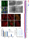
Protocol for myogenic differentiation. (A) Time line and phase contrast images of cells differentiated from human induced pluripotent stem cells (hiPSCs) at Day 0 (D0), D6, D8, D12, D17 and D30, corresponding to changes in the composition of the medium. Thiazovivin, an inhibitor of the ROCK pathway, was added at the time of plating and two days after. The composition of the medium is indicated for the different culture steps. CHIR: CHIR99021; LDN: LDN193189; F: FGF2; I: IGF1; H: HGF; DAPT: N-[N-(3,5-Difluorophenacetyl)-L-alanyl]-S-phenylglycine t-butyl ester. Cells could be frozen between Days 10 and 12 for storage, expansion and further differentiation. First contractions were visible between day 19 and 21 postdifferentiation. (B) Representative Z stack after confocal microscopy of cultures stained with anti-PAX3 antibody at Day 6 and Day 8 postdifferentiation, and Day 12 with anti-PAX7. Representative immunostaining of early myofibers at Day 14 stained for the sarcomeric Desmin (green). Cells were counterstained with DAPI.
Figure 1
Figure 1
Protocol for myogenic differentiation. (A) Time line and phase contrast images of cells differentiated from human induced pluripotent stem cells (hiPSCs) at Day 0 (D0), D6, D8, D12, D17 and D30, corresponding to changes in the composition of the medium. Thiazovivin, an inhibitor of the ROCK pathway, was added at the time of plating and two days after. The composition of the medium is indicated for the different culture steps. CHIR: CHIR99021; LDN: LDN193189; F: FGF2; I: IGF1; H: HGF; DAPT: N-[N-(3,5-Difluorophenacetyl)-L-alanyl]-S-phenylglycine t-butyl ester. Cells could be frozen between Days 10 and 12 for storage, expansion and further differentiation. First contractions were visible between day 19 and 21 postdifferentiation. (B) Representative Z stack after confocal microscopy of cultures stained with anti-PAX3 antibody at Day 6 and Day 8 postdifferentiation, and Day 12 with anti-PAX7. Representative immunostaining of early myofibers at Day 14 stained for the sarcomeric Desmin (green). Cells were counterstained with DAPI.
Figure 2
Figure 2
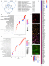
Ultrastructural and functional validation of hiPSC-derived myofibers. (A) Representative Z stacks of muscle fibers at Day 30, stained for Titin (TTN, green, upper panel) and merged between Desmin (DES, orange) and Titin filament (green, lower panel) at Day 30 postdifferentiation. Nuclei were counterstained with DAPI. (B) Ultrastructural features of differentiation. Control cells showed complete sarcomeric organization with highly organized myofibrillar patterns and fully mature sarcomeric banding pattern with clearly visible Z-lines (arrows, panels 2; 4), nuclei localized at the periphery of the fibers (panel 1, N) and large mitochondria (panel 3, M). Thick filaments were assembled with the formation of Z and M lines across the Myosin filaments (Panel 4). Z-lines were aligned, forming a clearly visible structural pattern (panels 1;2;4). We also observed I-bands of Actin filaments and A-bands (panels 2; 4). (C) Representative Z stacks of cultures stained for acetylcholine receptors (AChR) using Alpha Bungarotoxin coupled with Alexa 555 (White), and immunostaining of muscle fibers using anti-Titin (TTN, red), anti-Dystrophin (DYS, red), anti-Desmin (orange) and anti-NeuroFilament (NF, green) antibodies. We observed several undifferentiated acetylcholine receptors clusters at the surface of the myofibers (white). (D) Contractions of muscle fibers were recorded in real time by video microscopy in control cells (Supplementary Movie 1) prior to or after the addition of Alpha Bungarotoxin (α BTX) or Tetrodotoxin (TTX), to the cell medium, or after medium replacement and drug removal. Histogram displays the mean number of contractions ± S.D. shown by error bars, prior to drug addition (T0), 5 h (T 5H) or 24 h after (T 24 H), or 24 h after drug removal and medium replacement (T 24H washout). The addition of both drugs irreversibly inhibited cell contraction. (E) Heatmap of DEGs corresponding to the “contraction” GO terms in D8 vs. D17 and D17 vs. D30. Brackets identify genes differentially expressed for the different time points. Group 1, genes activated at D17; group 2, genes activated at D17 and D30; and group 3, genes upregulated at D8. (F) Intracellular calcium signaling in hiPSC-derived myofibers was measured using an Axio Observed microscope at day 30, day 120 (4 months) and day 150 (5 months) postdifferentiation. For each time point, >215 muscle fibers were analyzed separately. The number of contractions per minute and per fiber is reported as violin plots. No difference was observed in the calcium influx at day 120 and 150 compared to day 30 (ANOVA multiple comparison test followed by Brown-Forsythe and Welch correction p = 0.264 and p = 0.299; respectively). hiPSC-derived myofibers by transmission electron microscopy (TEM) at Day 45 post.
Figure 3
Figure 3
Transcriptomic profiling of in vitro differentiated innervated muscle fibers at day 8, 17 and 30 postdifferentiation. (A) Venn diagram showing the number of genes differentially regulated between D8 and D30, D8 and D17 or D17 and D30. The three time points correspond to the main medium changes. Genes were selected based on a padj <0.05. Lists of genes that are differentially expressed between D8–D17, D17–D30 and D8–30 are given in Supplementary Table S1. (B) List of the most significant Biological processes (BP) corresponding to the 302 genes common to the three time points with associated GO term and padj values. (C) Biological pathways significantly overrepresented between D8 and D17. (D) Representative Z stacks of hiPSC-derived motor neurons using anti HB9 antibodies (upper panel, red), anti-Neurofilament (NF, orange, medium) and merged (bottom) with nuclei counterstained with DAPI. (E) Immunostaining of hiPSC-derived motor neurons using anti HB9 antibodies (red) and anti-TUBβ3 (green) with nuclei counterstained with DAPI. (F) Biological pathways significantly overrepresented between D17 and D30. (G) Heatmap of the different myogenic transcription factors at D8, D17 and D30. (H) Heatmap of differentially expressed genes corresponding to “skeletal muscle differentiation” between D8, D17 and D30.
Figure 4
Figure 4
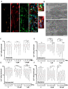
Characterization of NMJs and evaluation of the functional response to pharmaceutical drugs. (A) Representative Z stacks of cells stained at Day 30 postdifferentiation using alpha Bungarotoxin (aBTX, orange), anti-MyHC (MF20, red) or anti-S100β (green). NMJs are indicated by white arrows on the merge. The formation of functional NMJs was confirmed by the presence of S100β-positive cells expressed by Schwann cells capping the neuronal terminals. A higher magnification of different aBTX-positive NMJs is shown. (B) Transmission electron microscopy showing the presence of an axon positioned close to a muscle fiber. The basal lamina is visible (panels 1–2), as well as invagination of the plasma membrane (panel 2). (CF) We measured the transient calcium influx in control myofibers prior to drug addition (0 h) and at different time points after a single addition of different concentrations of each drug. Violin plots display the number of influxes per minute and per fiber (n > 50). Statistical significance was determined using an ANOVA multiple comparison test followed by Brown-Forsythe and Welch correction and comparison to the basal condition (prior to drug addition). C. 0; 2 and 20 μM for glutamate after 0; 10; 20; 30 min of incubation, (D) 0; 1 and 10 μM for Salbutamol after 0, 1, 2, 4 and 24 h of incubation. (E) 0, 0.5 and 1 mM for Carisoprodol after 0, 1, 2, 4 and 24 h of incubation. F. 0, 10 and 100 μM for Mexiletine after 0, 1, 2, 4 and 24 h of incubation.
Figure 5
Figure 5
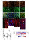
Structural and functional phenotyping of hiPSC-derived muscle cells from patients affected by muscular dystrophy. (A) Representative Z stacks of myofibers at day 45 with staining of sarcomeric Desmin (DES, orange) and Titin filament (TTN, green) for Duchenne Muscular Dystrophy (DMD), myotonic dystrophy (DM1), Facio Scapulo Humeral Dystrophy (FSHD) and Limb Girdle Muscular Dystrophy Type 2A (LGMD2A). (B) Representative Z stacks of cultures stained for acetylcholine receptors (AChR) using Alpha Bungarotoxin coupled with Alexa 555 (White, arrows) at the surface of hiPSC-derived muscle fiber stained in red using anti-Titin (TTN, red). Antineurofilament (NF, green) was used to visualize motor neurons axons. Nuclei were counterstained with DAPI. As in the control cells, we observed the presence of several undifferentiated acetylcholine receptors clusters at the surface of the myofibers for the different pathologies. (C) We compared the muscle fiber section of the Titin-stained fibers in control cells and the four different pathologies (DMD, DM1, FSHD or LGMD2A). For each condition, 500 fibers were analyzed, and the section of the fiber was determined at three different positions along each. Box plots display the mean sizes for the different samples. Values were compared using an ANOVA multiple comparison test, followed by Brown-Forsythe and Welch correction tests; ****p < 0.0001; ***p < 0.001; **p < 0.01. –(D) Heatmap for expression of different muscle and motor neuron markers over time for cells differentiated from the control (CTRL) or pathological (DMD, DM1, FSHD, LGMD2A) hiPSCs at D30 postdifferentiation, determined by RT-qPCR on biological triplicates, each realized in biological duplicates (n = 6 per data point). The names of the different genes are indicated on the bottom. Data for additional time points (D6, D8, D12, D17 and D21) are presented in the Supplementary Information section.
Figure 6
Figure 6
Functional and ultrastructural characterization of diseased hiPSC-derived muscle fibers. (A) Ultrastructural features of hiPSC-derived muscle fibers for different neuromuscular disorders. Electron microscopy was performed 60 days postdifferentiation. Representative images for DMD, DM1, FSHD and LGMD2A are presented. (B) Intracellular calcium signaling in hiPSC-derived myofibers was measured using an Axio Observed microscope at day 45 postdifferentiation. For each time point, >180 muscle fibers were analyzed. We report the number of influxes recorded per minute and per fiber as violin plots. Data were compared using an ANOVA multiple comparison test followed by Brown-Forsythe and Welch correction. DM1 myofibers presented lower calcium influx compared to the control (****; p < 0.0001), whereas calcium influx increased in FSHD and DMD myofibers (****; p < 0.0001). No difference was observed between the control and LGMD2A myofibers (p = 0.89).

References

    1. Ortiz-Vitali J.L., Darabi R. Ipscs as a platform for disease modeling, drug screening, and personalized therapy in muscular dystrophies. Cells. 2019;8:20. doi: 10.3390/cells8010020. - DOI - PMC - PubMed
    1. Darabi R., Gehlbach K., Bachoo R.M., Kamath S., Osawa M., Kamm K.E., Kyba M., Perlingeiro R.C. Functional skeletal muscle regeneration from differentiating embryonic stem cells. Nat. Med. 2008;14:134–143. doi: 10.1038/nm1705. - DOI - PubMed
    1. Maffioletti S.M., Gerli M.F., Ragazzi M., Dastidar S., Benedetti S., Loperfido M., VandenDriessche T., Chuah M.K., Tedesco F.S. Efficient derivation and inducible differentiation of expandable skeletal myogenic cells from human es and patient-specific ips cells. Nat. Protoc. 2015;10:941–958. doi: 10.1038/nprot.2015.057. - DOI - PubMed
    1. Pawlowski M., Ortmann D., Bertero A., Tavares J.M., Pedersen R.A., Vallier L., Kotter M.R.N. Inducible and deterministic forward programming of human pluripotent stem cells into neurons, skeletal myocytes, and oligodendrocytes. Stem Cell Rep. 2017;8:803–812. doi: 10.1016/j.stemcr.2017.02.016. - DOI - PMC - PubMed
    1. Barberi T., Bradbury M., Dincer Z., Panagiotakos G., Socci N.D., Studer L. Derivation of engraftable skeletal myoblasts from human embryonic stem cells. Nat. Med. 2007;13:642–648. doi: 10.1038/nm1533. - DOI - PubMed

Publication types

MeSH terms