. 2020 Jun 23;21(12):4467.
doi: 10.3390/ijms21124467.

SpeS: A Novel Superantigen and Its Potential as a Vaccine Adjuvant against Strangles

Affiliations

SpeS: A Novel Superantigen and Its Potential as a Vaccine Adjuvant against Strangles

C Coral Dominguez-Medina et al. Int J Mol Sci. .

Abstract

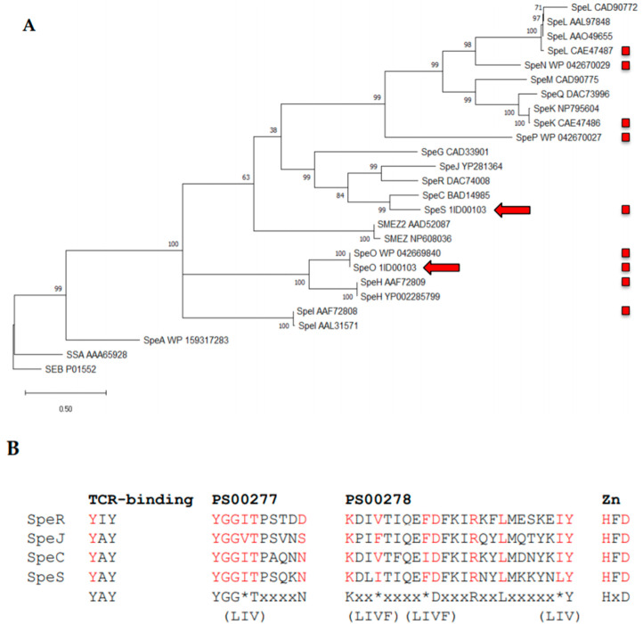
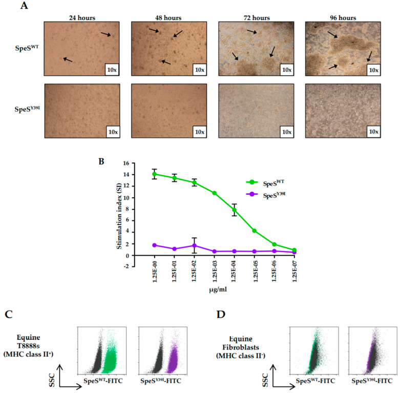
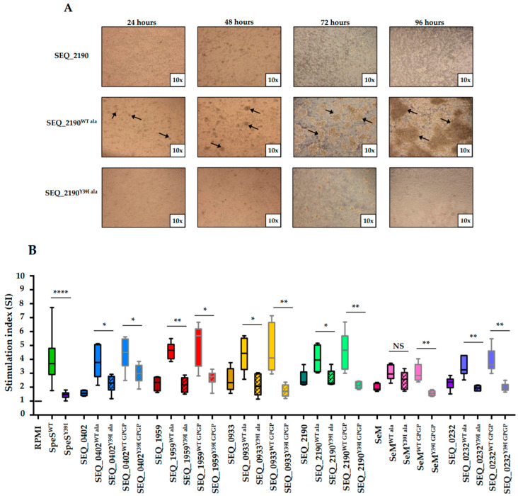
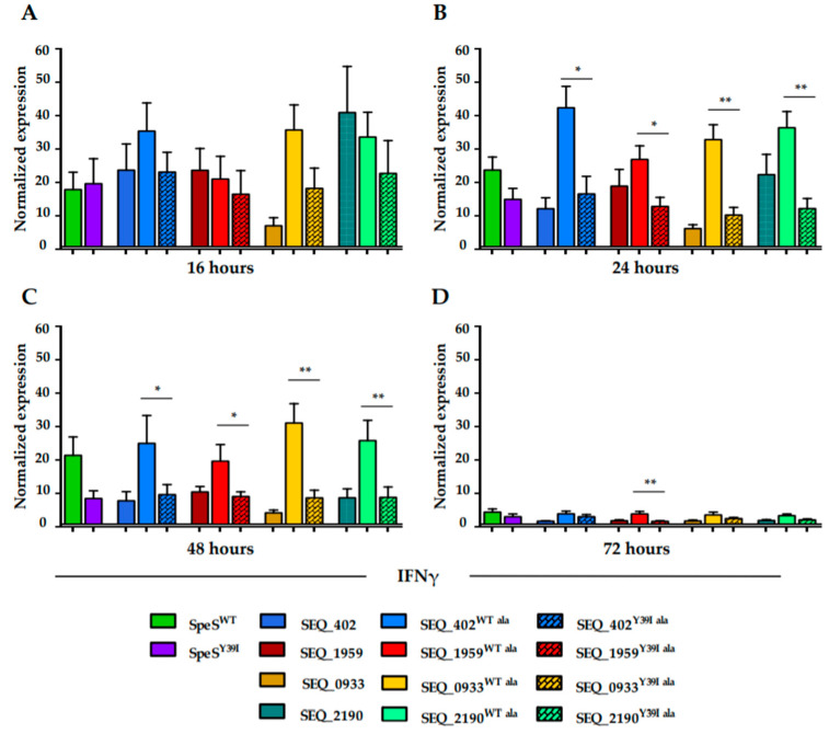
Bacterial superantigens (sAgs) are powerful activators of the immune response that trigger unspecific T cell responses accompanied by the release of proinflammatory cytokines. Streptococcus equi (S. equi) and Streptococcus zooepidemicus (S. zooepidemicus) produce sAgs that play an important role in their ability to cause disease. Strangles, caused by S. equi, is one of the most common infectious diseases of horses worldwide. Here, we report the identification of a new sAg of S. zooepidemicus, SpeS, and show that mutation of the putative T cell receptor (TCR)-binding motif (YAY to IAY) abrogated TCR-binding, whilst maintaining interaction with major histocompatibility complex (MHC) class II molecules. The fusion of SpeS and SpeSY39I to six S. equi surface proteins using two different peptide linkers was conducted to determine if MHC class II-binding properties were maintained. Proliferation assays, qPCR and flow cytometry analysis showed that SpeSY39I and its fusion proteins induced less mitogenic activity and interferon gamma expression when compared to SpeS, whilst retaining Antigen-Presenting Cell (APC)-binding properties. Our data suggest that SpeSY39I-surface protein fusions could be used to direct vaccine antigens towards antigen-presenting cells in vivo with the potential to enhance antigen presentation and improve immune responses.

Keywords: Streptococcus equi; Streptococcus zooepidemicus; adjuvant; horse; immune response; strangles; superantigen; vaccine.


Conflict of interest statement

The authors declare no competing interests.

Figures

Figure 1
Figure 1
Similarity of the novel superantigen SpeS to known superantigens. (A) The amino acid sequences of representatives of the 16 known streptococcal superantigens, SpeS and the staphylococcal superantigen SEB were obtained. Nomenclature has been standardized in accordance with Commons et al., 2014 [13]. The signal sequences were identified using SignalP [21] and removed. The remaining predicted amino acid sequences were utilized to generate a maximum likelihood tree in MEGA X [22], which was rooted to SEB. The percentage of trees in which the associated taxa clustered together is shown next to the branches. The tree is drawn to scale, with branch lengths measured in the number of substitutions per site. The red arrows indicate the location of the SpeO and SpeS sequences encoded within the Sz1ID00103 genome. Red boxes indicate superantigens encoded by S. equi or S. zooepidemicus. (B) Identification of signature domains found in superantigens. The amino acid sequence of SpeS was compared to the closest relatives SpeC, SpeJ and SpeR to identify motifs typically found in superantigens. The T cell receptor ß-binding motif YAY of SpeC [19,20], the consensus sequence Y-G-G-(LIV)-T-x4-N and K-x2-(LIVF)-x4-(LIVF)-D-x3-R-x2-L-x5-(LIV)-Y of the superantigen Prosite domains PS00277 and PS00278, respectively, and the zinc binding domain, HxD, were fully conserved in SpeS. Consensus matching residues are indicated in red. * represents one of the three or four possible amino acids indicated in brackets on the line directly below.
Figure 2
Figure 2
SpeSY39I does not stimulate T cell proliferation but maintains its ability to bind major histocompatibility complex (MHC) class II molecules in vitro. (A) Images of equine peripheral blood mononuclear cells (PBMCs) stimulated with 1 μg/mL of SpeSWT or SpeSY39I for 24, 48, 72 and 96 h. Cells were captured with a camera at 10× magnification. Pointed arrows indicate the formation of clusters of cells after stimulation with SpeSWT. (B) Dose response; a total of 1 × 10⁶ cells/mL PBMCs from two different donors were cultured in triplicate with 1/10 dilutions of SpeSWT or SpeSY39I for four days and incubated with 3HT for 16 h prior quantification of proliferating cells. Results are shown as stimulation index (SI) and error bars represent the standard error of the mean (SEM). (C,D) Flow cytometry analysis of SpeSWT (green) and SpeSY39I (purple) binding to MHC class II molecules. (C) Equine T8888s and (D) fibroblasts were incubated for 20 min at 4 °C with SpeSWT-Fluorescein isothiocyanate (FITC) or SpeSY39I-FITC in PBS, washed and the level of fluorescence was detected. Unstained cells (black) were used as a negative control.
Figure 3
Figure 3
S. equi surface proteins fused to SpeSY39I lack mitogenic activity in vitro. (A) Images of equine PBMCs cultured for 24, 48, 72 and 96 h with 1 μg/mL of SEQ_2190 or SEQ_2190 fused to SpeSWT or SpeSY39I. Images are representative of all the S. equi surface proteins and fusion proteins used. Photos of the cells were taken with a camera at 10× magnification. Pointed arrows show the clusters of cells formed after being exposed to the SpeSWT fusion protein. (B) Equine PBMCs (1 × 10⁶ cells/mL) from two different donors were stimulated in triplicate with 1 μg/mL of the S. equi surface proteins alone or as fusion proteins for four days. 3HT was added for 16 h before measuring the proliferation of cells. SpeSWT, SpeSY39I and medium alone were used as controls. Results are presented as SI. Data is from three independent experiments. Error bars correspond to SEM. * p ≤ 0.05, ** p ≤ 0.01, **** p ≤ 0.0001.
Figure 4
Figure 4
S. equi surface proteins fused to SpeSWT or SpeSY39I retain their ability to bind to MHC class II molecules in vitro. Flow cytometry analysis of SEQ_0402 (blue), SEQ_1959 (red), SEQ_0933 (yellow) and SEQ_2190 (aqua green) fusion proteins binding to MHC class II molecules. (A) Equine T8888s and (B) fibroblasts were incubated for 20 min at 4 °C with the FITC-labelled proteins, washed and the level of fluorescence analyzed. Unstained cells (black) were used as a negative control.
Figure 5
Figure 5
Levels of IFNγ (a proinflammatory cytokine) mRNA expression are reduced in PBMCs stimulated with SpeSY39I and S. equi surface proteins fused to SpeSY39I. A concentration of 4 × 10⁶ cells was cultured in duplicate with 1 μg/mL of PBS (control), SpeSWT, SpeSY39I or the fusion proteins for (A) 16 h, (B) 24 h, (C) 48 h and (D) 72 h. Samples were normalized against the housekeeping genes β-actin and GAPDH. Differences between the experimental samples and control samples were calculated. Results are from three different experiments where PBMCs from two different donors were used. Error bars represent SEM. * p ≤ 0.05, ** p ≤ 0.01.

Similar articles

Cited by

References

    1. Webb K., Jolley K.A., Mitchell Z., Robinson C., Newton J.R., Maiden M.C.J., Waller A. Development of an unambiguous and discriminatory multilocus sequence typing scheme for the Streptococcus zooepidemicus group. Microbiology. 2008;154:3016–3024. doi: 10.1099/mic.0.2008/018911-0. - DOI - PubMed
    1. Jorm L.R., Love D.N., Bailey G.D., McKay G.M., Briscoe D.A. Genetic structure of populations of beta-haemolytic Lancefield group C streptococci from horses and their association with disease. Res. Vet. Sci. 1994;57:292–299. doi: 10.1016/0034-5288(94)90120-1. - DOI - PubMed
    1. Waller A.S. Strangles: A pathogenic legacy of the war horse. Vet. Rec. 2016;178:91–92. doi: 10.1136/vr.i123. - DOI - PubMed
    1. Waller A.S. New perspectives for the diagnosis, control, treatment, and prevention of strangles in horses. Vet. Clin. North Am. Equine Pract. 2014;30:591–607. doi: 10.1016/j.cveq.2014.08.007. - DOI - PubMed
    1. Holden M.T., Heather Z., Paillot R., Steward K.F., Webb K., Ainslie F., Jourdan T., Bason N.C., Holroyd N.E., Mungall K., et al. Genomic evidence for the evolution of Streptococcus equi: Host restriction, increased virulence, and genetic exchange with human pathogens. PloS Pathog. 2009;5:e1000346. doi: 10.1371/journal.ppat.1000346. - DOI - PMC - PubMed

MeSH terms