. 2020 Jun 23;25(12):2890.
doi: 10.3390/molecules25122890.

Gold Nanoparticles as Colorimetric Sensors for the Detection of DNA Bases and Related Compounds

Affiliations

Gold Nanoparticles as Colorimetric Sensors for the Detection of DNA Bases and Related Compounds

Emilia Iglesias. Molecules. .

Abstract

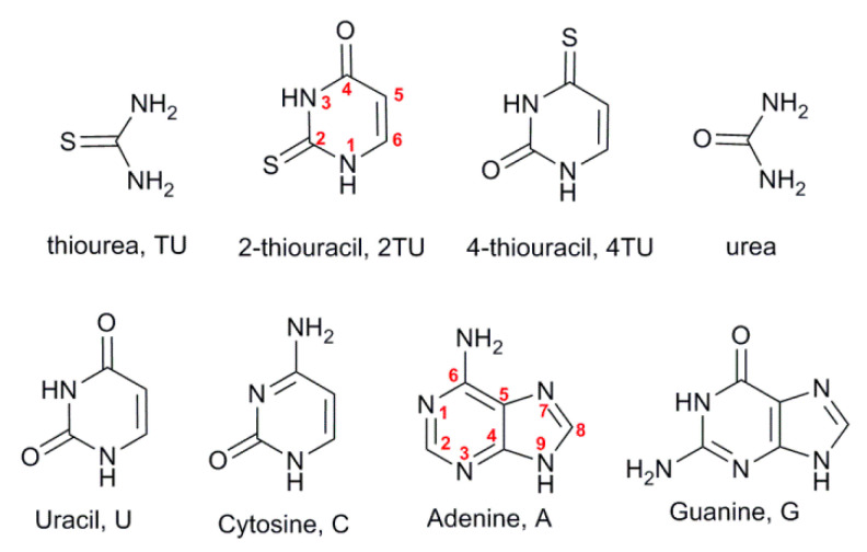
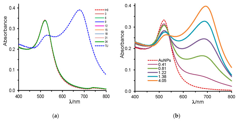
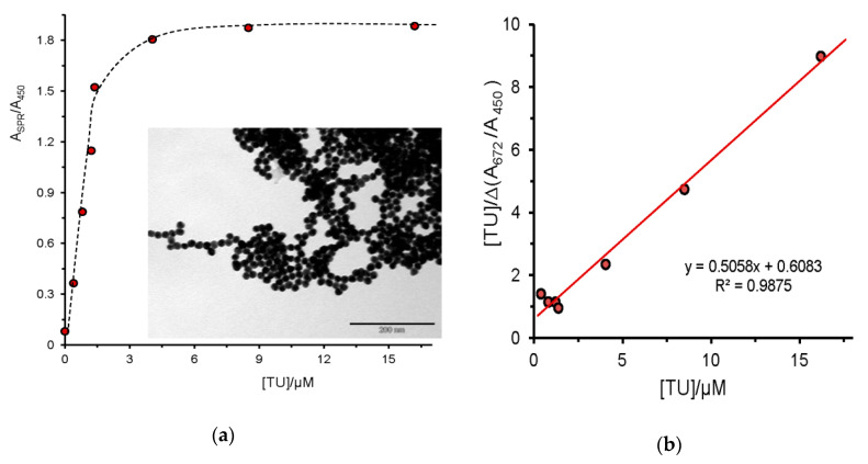
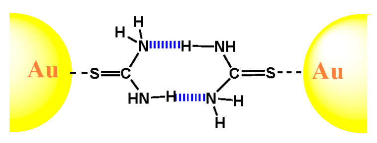
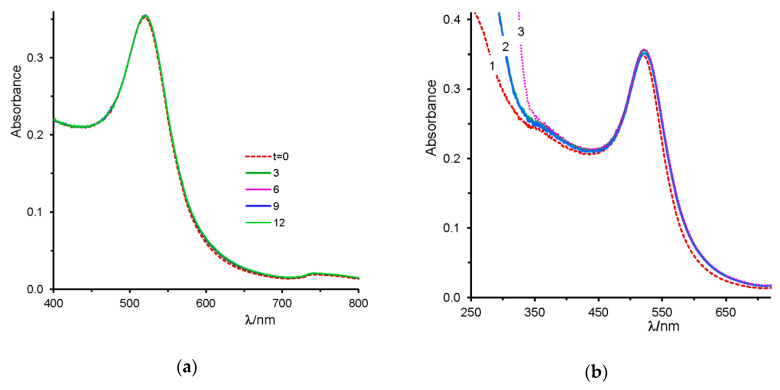
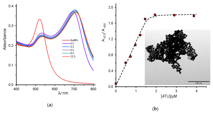
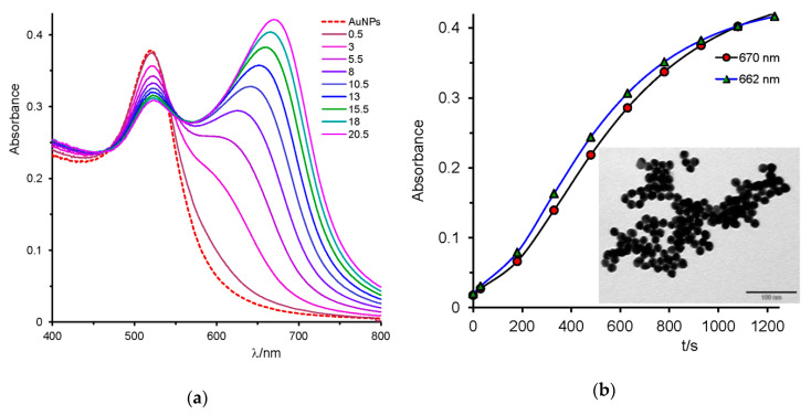
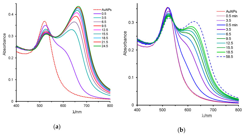
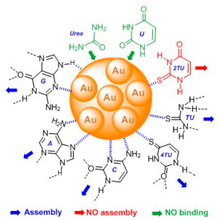
Results regarding interaction of colloidal gold solutions with nucleobases, including uracil (U), as well as its sulfur derivatives, 2-thiouracil (2TU) and 4-thiouracil (4TU), cytosine (C), adenine (A), and guanine (G), as well as urea and thiourea (TU), are reported. Anionic stabilized citrate gold nanoparticles (AuNPs) were synthesized by reducing the tetrachloroaurate (III) trihydrate with trisodium citrate. The surface plasmon resonance (SPR) band was used in the characterization of synthesized AuNPs, as well as transmission electron microscope (TEM) imaging, which was used in the characterization of dispersed and aggregated gold nanoparticles. Interactions of nucleobases with the gold surface was analyzed by following the plasmon absorbance band red shift of the AuNPs. The sulfur-containing compounds adsorbed to the nanoparticle surfaces by chemisorption-type interactions; with TU and 4TU, the process is accompanied by a sudden change in color; in contrast, 2TU forms stable functionalized gold nanoparticles. Urea and U do not adsorb to nanoparticle surfaces, but the other heterocyclic bases containing nitrogen interact effectively with the gold surface, causing the assembly of nanoparticles, even though the interparticle self-aggregation process was slower than that mediated by either TU or 4TU. The method is efficient in the colorimetric detection of nucleobases and derivatives at concentration levels on the order of 1 µM.

Keywords: biosensors; colorimetric detection; gold nanoparticles; nucleobases.


Conflict of interest statement

The authors declare no conflict of interest.

Figures

Scheme 1
Scheme 1
Molecular structures of the studied compounds.
Figure 1
Figure 1
(a) Visible spectrum of 0.78 nM gold nanoparticles (AuNPs; dashed red line), and in the presence of 2.4 mM urea recorded at 3 min intervals over a 24 min period; the dashed blue line shows the spectra after adding 30 µL of thiourea solution to get [TU] = 8.1 µM; (b) effect of TU concentration (see insert: [TU] = 0.41; 0.81; 1.22; 1.38, and 4.05 μM) on the surface plasmon resonance (SPR) band of AuNPs.
Figure 2
Figure 2
(a) Variation of the ratio of absorbance readings at 672 nm and 450 nm as a function of [TU], the insert shows the TEM image of AuNPs in the presence of 8.4 μM thiourea at 200 nm scale; (b) modified Benesi–Hildebrand plot for the absorbance ratio measured at A672/A450 as a function of TU concentration ([TU] = 0.4 to 16 μM).
Scheme 2
Scheme 2
Cartoon to illustrate the TU binding mode to a gold surface and H-bonding interactions between two adjacent nanoparticles.
Figure 3
Figure 3
Spectrum of AuNPs solution (dashed line) in the presence of (a) 0.196 mM uracil recorded over 12 min at 3 min intervals, and of (b) 9.34 µM 2-thiouracil (curve 2 repeated for six cycles at 3.5 min intervals) and 156 µM (curve 3, which matches curve 2 above 450 nm).
Figure 4
Figure 4
(a) Spectrum of AuNPs dispersion (dashed line) in the presence of 25.3 µM 4-thiouracil, recorded over 12.5 min at 3 min intervals; (b) ratio of the absorbance readings at 672 and 450 nm as a function of [4TU]; dashed line to guide the eye; the insert shows the typical TEM image of 4TU-mediated assembly of AuNPs.
Figure 5
Figure 5
(a) Evolution of the SPR band of gold nanoparticles in the presence of 11.3 µM cytosine recorded over 20.5 min at 2.5 min intervals (dashed spectrum is only NPs); (b) plot of the absorbance readings at (●) 670 nm and (▲) 662 nm as a function of time; the insert shown a TEM image of gold nanoparticles assembly mediated by cytosine.
Figure 6
Figure 6
Evolution spectra of the SPR band of AuNPs in the presence of (a) 1.55 µM adenine recorded over 24.5 min at 3 min intervals; (dashed curve) only nanoparticles (NPs); (b) dotted curves correspond to AuNPs in the absence and in the presence of [Mn+2] = 0.046 mM after 3.5 min; solid curves were recorded at 3 min intervals after adding 8.4 µM guanine hydrochloride; and dashed curves are 40 min after the latter.
Scheme 3
Scheme 3
Schematic representation of the binding modes of the studied DNA bases and related compounds: Black structures represent the bases that mediate AuNPs aggregation; the red compound coats the gold surface, but does not induce aggregation, and green compounds neither bind to the gold surface nor induce NPs assembly.

Similar articles

Cited by

References

    1. Liu B., Liu J. Interface-driven hybrid materials based on DNA-functionalized gold nanoparticles. Matter. 2019;1:825–847. doi: 10.1016/j.matt.2019.08.008. - DOI
    1. Carnerero J.M., Jimenez-Ruiz A., Castilli P.M., Prado-Gotor R. Covalent and non-covalent DNA-gold nanoparticle interactions: New avenues of research. ChemPhysChem. 2017;18:17–33. doi: 10.1002/cphc.201601077. - DOI - PubMed
    1. Montané X., Bajek A., Roszkowski K., Montornés J.M., Giamberini M., Roszkowski S., Kowalczyk O., Garcia-Valls R., Tylkowski B. Encapsulation for Cancer Therapy. Molecules. 2020;25:1605. doi: 10.3390/molecules25071605. - DOI - PMC - PubMed
    1. Jans H., Huo Q. Gold nanoparticle-enabled biological and chemical detection analysis. Chem. Soc. Rev. 2012;41:2849–2866. doi: 10.1039/C1CS15280G. - DOI - PubMed
    1. Saka K., Agasti S.S., Kim C., Li X., Rotello V.M. Gold nanoparticles in chemical and biological sensing. Chem. Rev. 2012;112:2739–2779. - PMC - PubMed

LinkOut - more resources