. 2020:159:1-22.
doi: 10.1016/bs.mcb.2020.04.009. Epub 2020 May 13.

Generation of esophageal organoids and organotypic raft cultures from human pluripotent stem cells

Affiliations

Generation of esophageal organoids and organotypic raft cultures from human pluripotent stem cells

Vered Shacham-Silverberg et al. Methods Cell Biol. 2020.

Abstract

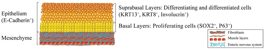
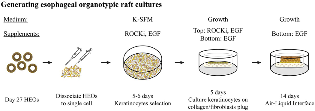
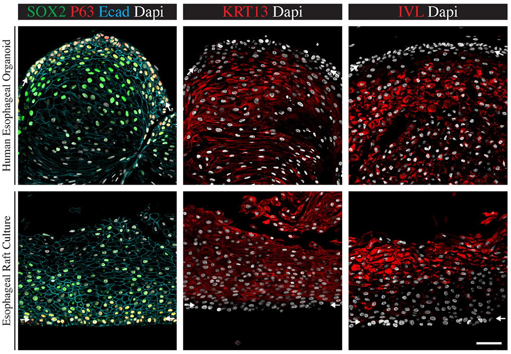
The human and murine esophagus have some substantial differences that limit the utility of mouse as a model to study human esophagus development and disease. Due to these limitations several recent reports describe the development of methods to generate human esophageal tissues via the directed differentiation of pluripotent stem cells. Methods for differentiation are based on knowledge of years of studying embryonic development of the esophagus in vertebrate animal models. Esophageal tissues derived from human pluripotent stem cells have been used to study both development and diseases affecting the esophagus. Here, we provide a detailed protocol for the directed differentiation of human pluripotent stem cells into human esophageal organoids and organotypic raft cultures, that are highly similar, morphologically and transcriptionally, to the human esophagus epithelium. We discuss limitations of the current esophageal models and the importance of engineering more complex tissue models with muscle and enteric nerves. Moving forward, these models might be utilized for the development of personalized treatments, as well as other therapeutic solutions.

Keywords: Anterior foregut; Definitive endoderm; Esophageal organoids; Esophageal raft cultures; Human pluripotent stem cells.


Figures

FIG. 1
FIG. 1
The human esophagus is consisting of mesenchyme and epithelium. The human esophagus mesenchyme is mesoderm-derived and is consists of fibroblasts and several muscle layers, which are innervated by the enteric nervous system, which is neural crest-derived. The esophagus epithelium is endoderm-derived and is a stratified squamous epithelium, expressing the epithelium marker E-Cadherin that consists of basal layers of proliferating cells, expressing SOX2 and P63 among other markers. Once the cells start to differentiate, they migrate toward the lumen and populate the suprabasal layers, expressing KRT13 and Involucrin, among other markers.
FIG. 2
FIG. 2
Protocol for the generation of human esophageal organoids. (A) hPSC are directly differentiated into definitive endoderm (day 3) and then into anterior foregut spheroids (day 6) which are then embedded in 3D matrix where differentiation and maturation of the spheroids continues for the generation of human esophageal organoids (HEOs). HEOs can survive up to 2 months in vitro, from day of embedding, by reducing organoids density 21 days after embedding (Day 27). Media and supplements are listed above each stage. For complete medium composition and supplements concentrations turn to Section 5.2 in the text. (B) Bright field images representing different stages of differentiation protocol. B-6 is zoom-in on dashed rectangle in B-5. Dashed circle in B-7 marks 3D matrix bubble margin. B-1, 5, 6, 7 and 8 were taken in up-right stereomicroscope (Leica, cat. # S8APO) scale bar: 500 μm. B-2, 3 and 4 were taken in inverted microscope (Nikon, TMS model), 10× magnification, scale bar—100 μm.
FIG. 3
FIG. 3
Generation of organotypic raft cultures from HEOs. Day 27 HEOs (21 days post embedding) are collected into a conical tube and dissociated to single cell using enzyme-based dissociation and serial passaging through pipette tips and syringe needles and re-cultured as monolayer for 5–6 days for keratinocyte selection. Then cells are re-dissociated and cultured on top of a collagen/mice fibroblasts plugs in trans-well inserts. After 5 days of culture, medium is removed from top compartment to generate air-liquid interface (ALI). Cells continue to proliferate and differentiate for up to 2 weeks to generate esophageal organotypic raft cultures. For complete and detailed protocol and medium composition turn to Section 5.3 in the text.
FIG. 4
FIG. 4
Immunofluorescent analysis of HEOs and raft cultures. HEOs (Top panel) and organotypic raft cultures (Bottom panel) express esophagus epithelium specific markers. The entire epithelium is expressing the epithelium marker E-cadherin. Basal layers cells expressing P63 and SOX2 but lack the expression of keratin 13 (KRT13) and involucrin (IVL) as marked by white arrows. Suprabasal layers of differentiated cells express KRT13 and IVL but lack P63 expression. Scale bar—50 μm. Images where modified from Trisno, S. L., Philo, K. E. D., McCracken, K. W., Catá, E. M., Ruiz-Torres, S., Rankin, S. A., et al. (2018). Esophageal organoids from human pluripotent stem cells delineate Sox2 functions during esophageal specification. Cell Stem Cell, 23(4), 501–515.e7. doi: 10.1016/j.stem.2018.08.008 (Fig. 4), pseudo colors and brightness were altered from original images for visualization purposes.

Similar articles

Cited by

References

    1. Anacker D, & Moody C (2012). Generation of organotypic raft cultures from primary human keratinocytes. Journal of Visualized Experiments, (60), e3668. 10.3791/3668. - DOI - PMC - PubMed
    1. Bajpai R, Chen DA, Rada-Iglesias A, Zhang J, Xiong Y, Helms J, et al. (2010). CHD7 cooperates with PBAF to control multipotent neural crest formation. Nature, 463(7283), 958–962. 10.1038/nature08733. - DOI - PMC - PubMed
    1. D’Amour KA, Agulnick AD, Eliazer S, Kelly OG, Kroon E, & Baetge EE (2005). Efficient differentiation of human embryonic stem cells to definitive endoderm. Nature Biotechnology, 23(12), 1534–1541. 10.1038/nbt1163. - DOI - PubMed
    1. Fausett SR, & Klingensmith J (2012). Compartmentalization of the foregut tube: Developmental origins of the trachea and esophagus. Wiley Interdisciplinary Reviews: Developmental Biology, 1(2), 184–202. 10.1002/wdev.12. - DOI - PubMed
    1. Haeri H, Mardany O, Asadi-Amoli F, & Shahsiah R (2013). Human papilloma virus and esophageal squamous cell carcinoma. Acta Medica Iranica, 51 (4), 242–245. Retrieved from http://www.ncbi.nlm.nih.gov/pubmed/23690103. - PubMed

LinkOut - more resources