. 2020:159:201-227.
doi: 10.1016/bs.mcb.2020.03.001. Epub 2020 Apr 8.

Generation of human colonic organoids from human pluripotent stem cells

Affiliations

Generation of human colonic organoids from human pluripotent stem cells

Abdelkader Daoud et al. Methods Cell Biol. 2020.

Abstract

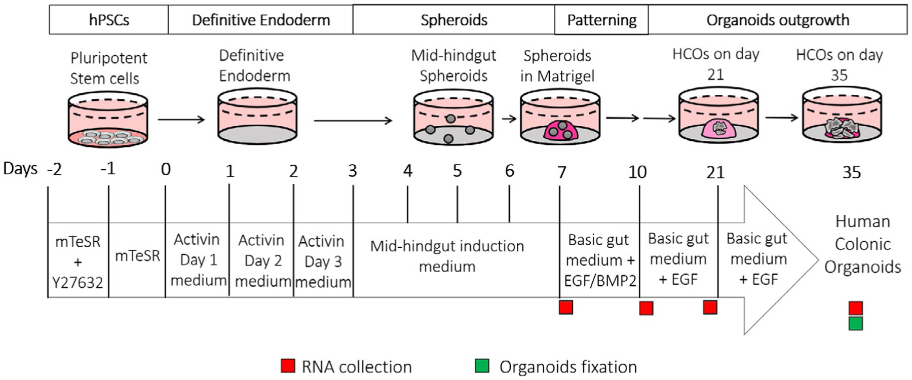
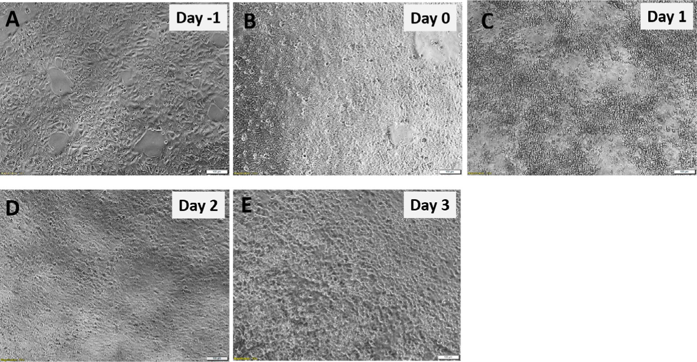
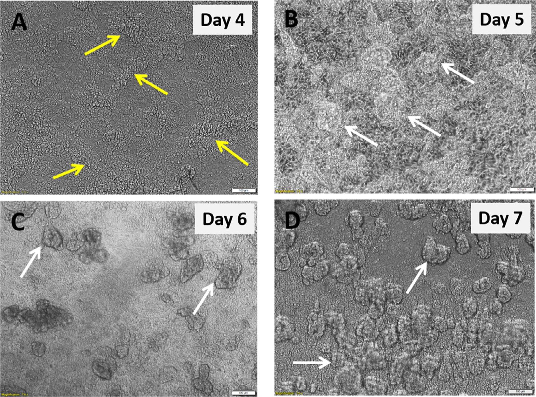
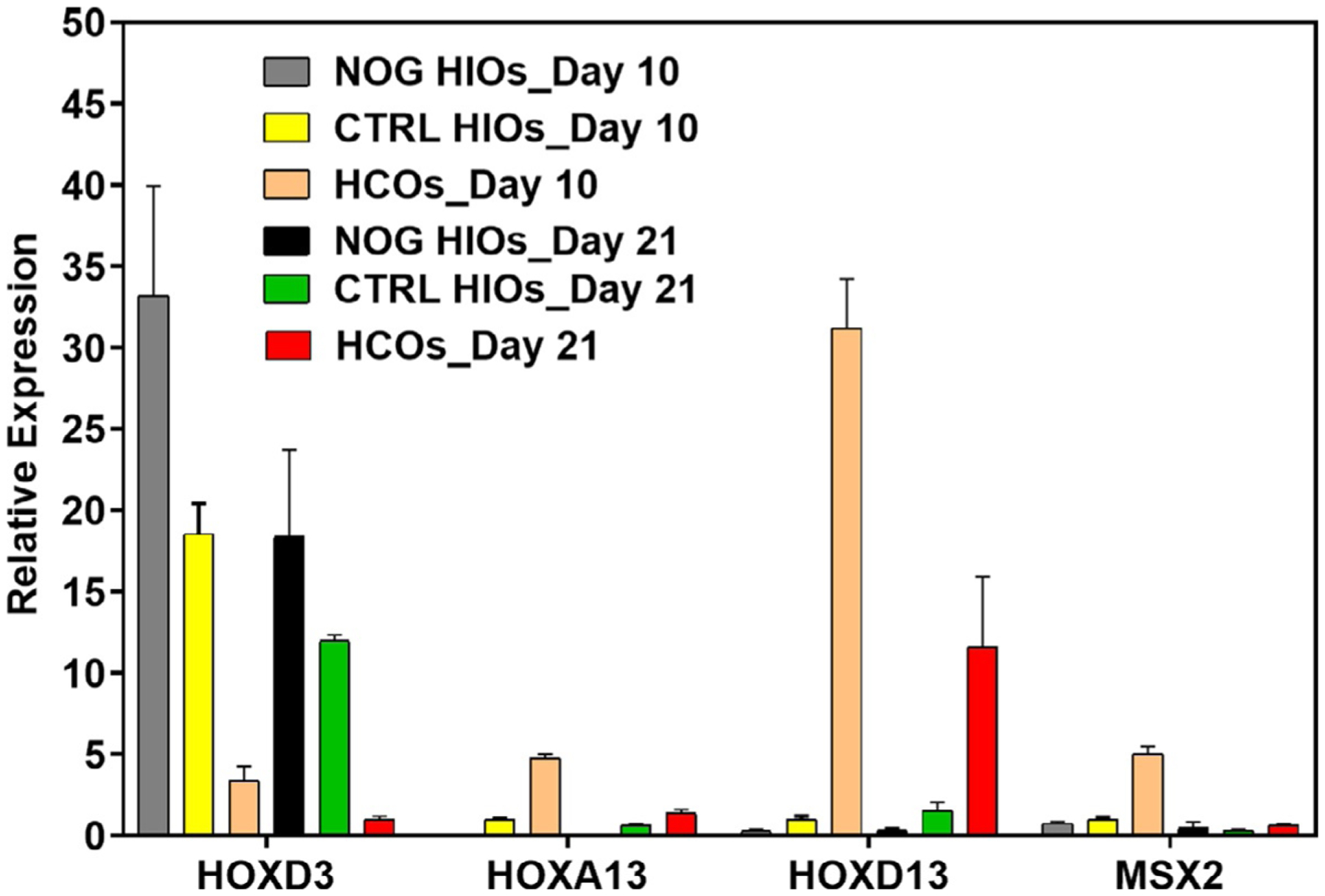
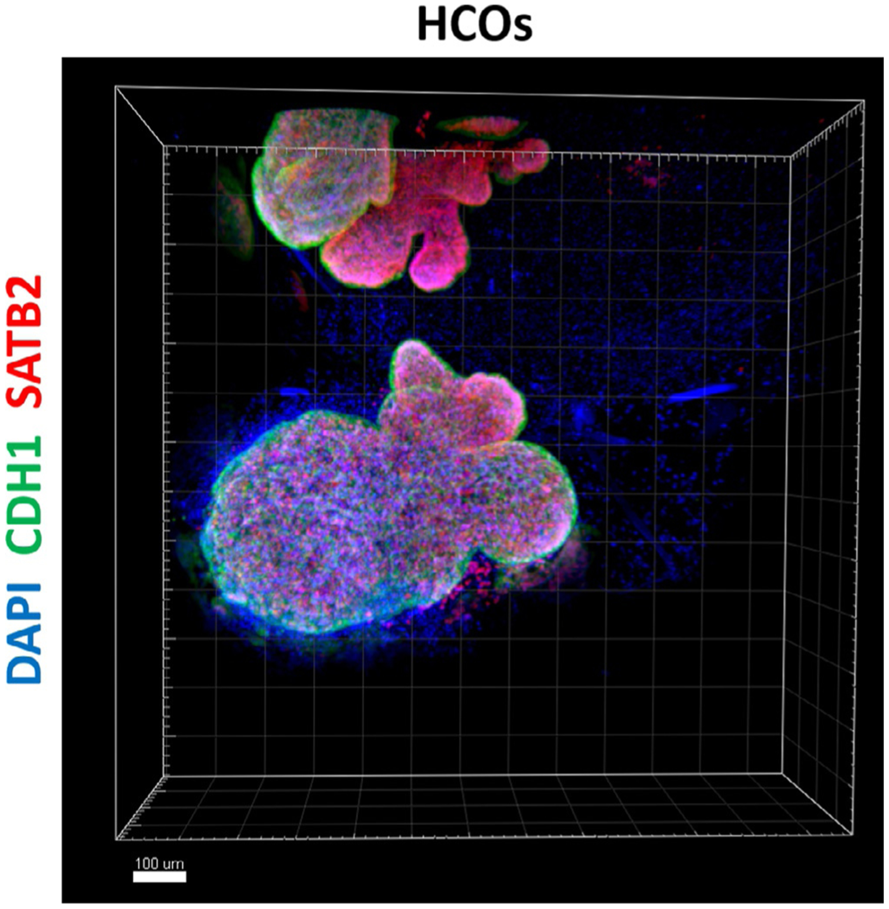
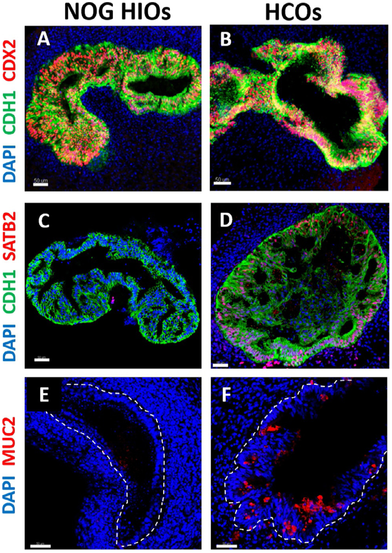
Advances in human pluripotent stem cell (hPSC) biology now allow the generation of organoids that resemble different regions of the gastrointestinal tract. Generation of region-specific organoids has been facilitated by developmental biology studies carried out in model organisms such as mouse, frog and chick. By mimicking embryonic development, hPSC-derived human colonic organoids (HCOs) can be generated through a stepwise differentiation, first into definitive endoderm (DE), then into mid/hindgut spheroids which are then patterned into posterior gut tissue which gives rise to HCOs following prolonged in vitro culture. HCOs undergo transitions similar to those observed in the developing colon of model organisms and human embryos. HCOs develop into tissue that resembles fetal colon on the basis of morphology, gene expression and presence of differentiated cell types. Generation of HCOs without the proper training or expertise can be a daunting task. Here, we describe a detailed protocol for differentiating hPSCs into HCOs, we include suggestions for troubleshooting these differentiations, and we discuss experimental design considerations. We have also highlighted the key advantages and limitations of the system.

Keywords: Definitive endoderm; Hindgut; Human colonic organoid; Human pluripotent stem cells.


Figures

FIG. 1
FIG. 1
Overview of the protocol for generating colonic organoids (HCOs) in vitro. The general timeline of HCO generation. Specific media for each step, are indicated; recommended timepoints for samples collection are also depicted by green (organoids) and red (RNA) squares.
FIG. 2
FIG. 2
Definitive endoderm (DE) monolayer generation from hPSCs. hPSCs morphology before (A and B) and after Activin A Day 1 (C) after Activin A Day 2 (D) and after Activin A Day 3 (E). Scale bar = 100μm.
FIG. 3
FIG. 3
Mid-hindgut spheroids generation from the definitive endoderm. DE monolayer morphology 24h after adding mid-hindgut induction (MHGI) medium Day 1 (A) MHGI medium Day 2 (B) MHGI medium Day 3 (C) and MHGI medium Day 4 (D). Yellow arrows indicate tubular structures and white arrows indicates mid-hindgut spheroids. Scale bar = 100μm.
FIG. 4
FIG. 4
Gene expression of HOX genes and MSX2 in human colonic and intestinal organoids. RNA is isolated from Noggin HIOs (NOG HIOs), CTRL organoids and HCOs on Day 10 and 21. qRT-PCR was performed as previously described in methods and relative expression of HOX genes and MSX2 was calculated for each group compared to control organoids of Day 10.
FIG. 5
FIG. 5
Whole mount immunostaining of human colonic organoid. Whole mount immunostaining of colonic marker SATB2 (red), CDH1 (green) and DAPI (blue) of 35-days old organoids that resulted from a 3-day treatment of gut tube spheroids with BMP2 (HCOs). Scale bar = 100μm.
FIG. 6
FIG. 6
Characterization of human intestinal and colonic organoids by immunostaining. (A and B) Immunofluorescence of intestinal epithelial marker CDX2 (red), epithelial marker CDH1 (green) and DAPI (blue) of 35-days old organoids that resulted from a 3-days treatment of gut tube spheroids with NOGGIN (NOG HIOs) or BMP2 (HCOs). (C and D) Immunostaining of colonic marker SATB2 (red), CDH1 (green) and DAPI (blue) of NOG HIOs and HCOs. (E and F) Immunofluorescence of goblet cells marker MUC2 (red) and DAPI (blue) of NOG HIOs and HCOs. The dotted lines in E and F denote the epithelium. Scale bar = 50μm.

Similar articles

Cited by

References

    1. Bartfeld S, Bayram T, van de Wetering M, Huch M, Begthel H, Kujala P, et al. (2015). In vitro expansion of human gastric epithelial stem cells and their responses to bacterial infection. Gastroenterology, 148(1), 126–136.e126. 10.1053/j.gastro.2014.09.042. - DOI - PMC - PubMed
    1. Crespo M, Vilar E, Tsai SY, Chang K, Amin S, Srinivasan T, et al. (2017). Colonic organoids derived from human induced pluripotent stem cells for modeling colorectal cancer and drug testing. Nature Medicine, 23(7), 878–884. 10.1038/nm.4355. - DOI - PMC - PubMed
    1. D’Amour KA, Agulnick AD, Eliazer S, Kelly OG, Kroon E, & Baetge EE (2005). Efficient differentiation of human embryonic stem cells to definitive endoderm. Nature Biotechnology, 23(12), 1534–1541. 10.1038/nbt1163. - DOI - PubMed
    1. Dye BR, Hill DR, Ferguson MA, Tsai YH, Nagy MS, Dyal R, et al. (2015). In vitro generation of human pluripotent stem cell derived lung organoids. eLife, 4, e05098. 10.7554/eLife.05098. - DOI - PMC - PubMed
    1. Fordham RP, Yui S, Hannan NR, Soendergaard C, Madgwick A, Schweiger PJ, et al. (2013). Transplantation of expanded fetal intestinal progenitors contributes to colon regeneration after injury. Cell Stem Cell, 13(6), 734–744. 10.1016/j.stem.2013.09.015. - DOI - PMC - PubMed

LinkOut - more resources