

Decreased salivary lactoferrin levels are specific to Alzheimer's disease
- PMID: 32586758
- PMCID: PMC7378957
- DOI: 10.1016/j.ebiom.2020.102834
Decreased salivary lactoferrin levels are specific to Alzheimer's disease
Abstract
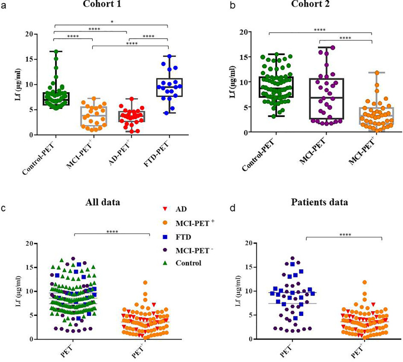
Background: Evidences of infectious pathogens in Alzheimer's disease (AD) brains may suggest a deteriorated innate immune system in AD pathophysiology. We previously demonstrated reduced salivary lactoferrin (Lf) levels, one of the major antimicrobial proteins, in AD patients.
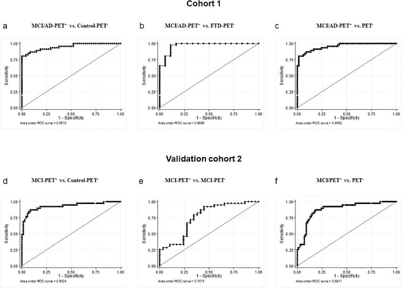
Methods: To assess the clinical utility of salivary Lf for AD diagnosis, we examine the relationship between salivary Lf and cerebral amyloid-β (Aβ) load using amyloid-Positron-Emission Tomography (PET) neuroimaging, in two different cross-sectional cohorts including patients with different neurodegenerative disorders.
Findings: The diagnostic performance of salivary Lf in the cohort 1 had an area under the curve [AUC] of 0•95 (0•911-0•992) for the differentiation of the prodromal AD/AD group positive for amyloid-PET (PET+) versus healthy group, and 0•97 (0•924-1) versus the frontotemporal dementia (FTD) group. In the cohort 2, salivary Lf had also an excellent diagnostic performance in the health control group versus prodromal AD comparison: AUC 0•93 (0•876-0•989). Salivary Lf detected prodromal AD and AD dementia distinguishing them from FTD with over 87% sensitivity and 91% specificity.
Interpretation: Salivary Lf seems to have a very good diagnostic performance to detect AD. Our findings support the possible utility of salivary Lf as a new non-invasive and cost-effective AD biomarker.
Funding: Instituto de Salud Carlos III (FIS15/00780, FIS18/00118), FEDER, Comunidad de Madrid (S2017/BMD-3700; NEUROMETAB-CM), and CIBERNED (PI2016/01) to E.C.; Spanish Ministry of Economy and Competitiveness (SAF2017-85310-R) to J.L.C., and (PSI2017-85311-P) to M.A.; International Centre on ageing CENIE-POCTEP (0348_CIE_6_E) to M.A.; Instituto de Salud Carlos III (PIE16/00021, PI17/01799), to H.B.
Keywords: Alzheimer´s disease; Biomarkers; Frontotemporal dementia; Lactoferrin; Pet imaging; Saliva.
Copyright © 2020 The Authors. Published by Elsevier B.V. All rights reserved.
Figures
Comment in
-
Examination of a non-invasive biomarker for the diagnosis of prodromal Alzheimer's disease and Alzheimer's disease Dementia.EBioMedicine. 2020 Jul;57:102882. doi: 10.1016/j.ebiom.2020.102882. Epub 2020 Jul 7. EBioMedicine. 2020. PMID: 32650274 Free PMC article. No abstract available.
References
-
- Lovheim H., Gilthorpe J., Adolfsson R., Nilsson L.G., Elgh F. Reactivated herpes simplex infection increases the risk of Alzheimer's disease. Alzheimer's Dement J Alzheimer's Assoc. 2015 Jun;11(6):593–599. - PubMed
-
- Lovheim H., Gilthorpe J., Johansson A., Eriksson S., Hallmans G., Elgh F. Herpes simplex infection and the risk of Alzheimer's disease: A nested case-control study. Alzheimer's Dement J Alzheimer's Assoc. 2015 Jun;11(6):587–592. - PubMed
MeSH terms
Substances
LinkOut - more resources
Full Text Sources
Other Literature Sources
Medical
Research Materials
