

Acute necrotizing encephalopathy with SARS-CoV-2 RNA confirmed in cerebrospinal fluid
- PMID: 32586897
- PMCID: PMC7538220
- DOI: 10.1212/WNL.0000000000010250
Acute necrotizing encephalopathy with SARS-CoV-2 RNA confirmed in cerebrospinal fluid
Abstract
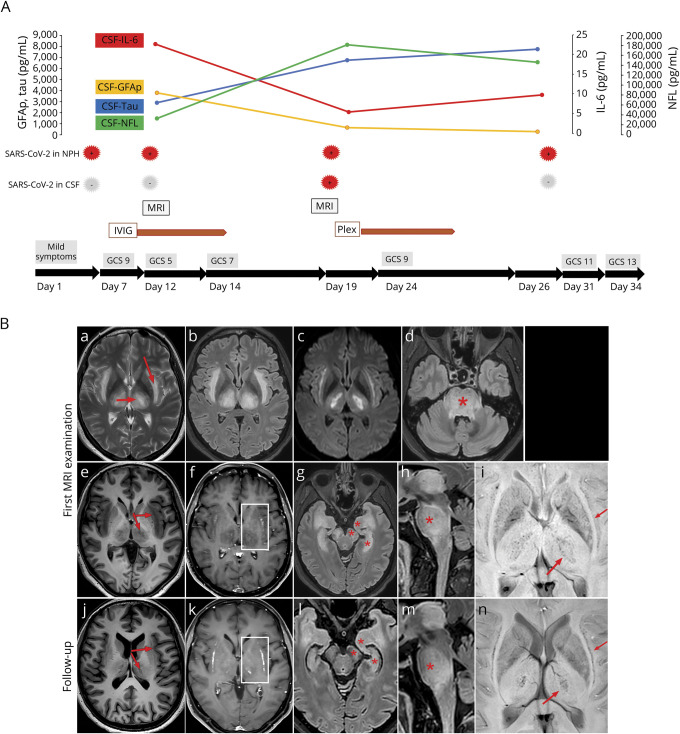
Here, we report a case of COVID-19-related acute necrotizing encephalopathy where SARS-CoV-2 RNA was found in CSF 19 days after symptom onset after testing negative twice. Although monocytes and protein levels in CSF were only marginally increased, and our patient never experienced a hyperinflammatory state, her neurologic function deteriorated into coma. MRI of the brain showed pathologic signal symmetrically in central thalami, subinsular regions, medial temporal lobes, and brain stem. Extremely high concentrations of the neuronal injury markers neurofilament light and tau, as well as an astrocytic activation marker, glial fibrillary acidic protein, were measured in CSF. Neuronal rescue proteins and other pathways were elevated in the in-depth proteomics analysis. The patient received IV immunoglobulins and plasma exchange. Her neurologic status improved, and she was extubated 4 weeks after symptom onset. This case report highlights the neurotropism of SARS-CoV-2 in selected patients and emphasizes the importance of repeated lumbar punctures and CSF analyses in patients with suspected COVID-19 and neurologic symptoms.
Copyright © 2020 The Author(s). Published by Wolters Kluwer Health, Inc. on behalf of the American Academy of Neurology.
Figures
Comment in
-
Editors' Note: Etiologic Uncertainties Regarding Neurologic Presentations Associated With COVID-19.Neurology. 2021 Aug 3;97(5):251. doi: 10.1212/WNL.0000000000012358. Neurology. 2021. PMID: 34341077 No abstract available.
-
Reader Response: Acute Necrotizing Encephalopathy With SARS-CoV-2 RNA Confirmed in Cerebrospinal Fluid.Neurology. 2021 Aug 3;97(5):252. doi: 10.1212/WNL.0000000000012357. Neurology. 2021. PMID: 34341078 No abstract available.
References
-
- Mizuguchi M. Acute necrotizing encephalopathy of childhood: a novel form of acute encephalopathy prevalent in Japan and Taiwan. Brain Dev 1997;19:81–92. - PubMed
Publication types
MeSH terms
Substances
LinkOut - more resources
Full Text Sources
Other Literature Sources
Medical
Miscellaneous