. 2020 Sep;41(9):1703-1706.
doi: 10.3174/ajnr.A6675. Epub 2020 Jun 25.

Anosmia in COVID-19 Associated with Injury to the Olfactory Bulbs Evident on MRI

Affiliations

Anosmia in COVID-19 Associated with Injury to the Olfactory Bulbs Evident on MRI

M F V V Aragão et al. AJNR Am J Neuroradiol. 2020 Sep.

Abstract

Patients with coronavirus disease 2019 (COVID-19) may have symptoms of anosmia or partial loss of the sense of smell, often accompanied by changes in taste. We report 5 cases (3 with anosmia) of adult patients with COVID-19 in whom injury to the olfactory bulbs was interpreted as microbleeding or abnormal enhancement on MR imaging. The patients had persistent headache (n = 4) or motor deficits (n = 1). This olfactory bulb injury may be the mechanism by which the Severe Acute Respiratory Syndrome coronavirus 2 causes olfactory dysfunction.


Figures

FIG 1.
FIG 1.
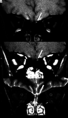
MR imaging shows probably microbleeding (methemoglobin) in the left olfactory bulb of a patient (case 1) with COVID-19 and anosmia. The left olfactory bulb (long arrows) has partial hyperintensity on precontrast fat-suppressed T1WI (A) and also on postcontrast fat-suppressed T1WI (B) and STIR (C).
FIG 2.
FIG 2.
The coronal postcontrast fat-suppressed T1WI shows hyperintensity suggestive of enhancement or methemoglobin in the olfactory bulbs of 4 patients with COVID-19 (A–D; cases 2–5) compared with a healthy patient with normal olfactory bulbs (E and F). The coronal postcontrast fat-suppressed T1WI in 3 patients with COVID-19 (A–C; cases 2–4) shows that both olfactory bulbs (long arrows) are small oval images that are hyperintense with contrast, having signal intensity higher than the intensity of the cortex. D, A patient (case 5) with COVID-19 shows hyperintensity only on the left bulb (long arrow), the right olfactory bulb being normal (short arrow). In a healthy 60-year-old man, the coronal T2WI (E) and the postcontrast fat-suppressed T1WI (F) demonstrate normal olfactory bulbs (long arrows), which are isointense to the cortex and normally hypointense on postgadolinium sequence (F).

Comment in

  • Reply.
    Aragão MFVV, Leal MC, Fonseca TM, Cartaxo Filho OQ, Valença MM. Aragão MFVV, et al. AJNR Am J Neuroradiol. 2021 Jan;42(2):E2-E3. doi: 10.3174/ajnr.A6943. Epub 2020 Dec 3. AJNR Am J Neuroradiol. 2021. PMID: 33272952 Free PMC article. No abstract available.
  • Seeing What We Expect to See in COVID-19.
    Mamourian A. Mamourian A. AJNR Am J Neuroradiol. 2021 Jan;42(2):E1. doi: 10.3174/ajnr.A6912. Epub 2020 Dec 3. AJNR Am J Neuroradiol. 2021. PMID: 33272953 Free PMC article. No abstract available.
  • Reply.
    Aragao MFVV, deAndrade PHP, da Nobrega Segundo VS, Marques MM, de Azevedo HJG, de Mello RJV, de Carvalho Leal M, Soares ML, Valenca MM. Aragao MFVV, et al. AJNR Am J Neuroradiol. 2021 Sep;42(9):E66-E68. doi: 10.3174/ajnr.A7234. Epub 2021 Aug 5. AJNR Am J Neuroradiol. 2021. PMID: 34353782 Free PMC article. No abstract available.
  • Susceptibility Artifacts in the Anterior Cranial Fossa Mimicking Hemorrhage in Patients with Anosmia.
    Manov JJ, Alfonso M, Saigal G. Manov JJ, et al. AJNR Am J Neuroradiol. 2021 Sep;42(9):E64-E65. doi: 10.3174/ajnr.A7184. Epub 2021 Aug 5. AJNR Am J Neuroradiol. 2021. PMID: 34353783 Free PMC article. No abstract available.

Similar articles

Cited by

References

    1. Fodoulian L, Tuberosa J, Rossier D, et al. . SARS-CoV-2 receptor and entry genes are expressed by sustentacular cells in the human olfactory neuroepithelium. bioRxiv 2020. https://www.biorxiv.org/content/10.1101/2020.03.31.013268v1. Accessed May 12, 2020 - DOI - PMC - PubMed
    1. Desforges M, Le Coupanec A, Dubeau P, et al. . Human coronaviruses and other respiratory viruses: underestimated opportunistic pathogens of the central nervous system? Viruses 2019;12:14 10.3390/v12010014 - DOI - PMC - PubMed
    1. Morris M, Zohrabian VM. Neuroradiologists, be mindful of the neuroinvasive potential of COVID-19. AJNR Am J Neuroradiol 2020. April 30. [Epub ahead of print] 10.3174/ajnr.A6551 - DOI - PMC - PubMed
    1. Montalvan V, Lee J, Bueso T, et al. . Neurological manifestations of COVID-19 and other coronavirus infections: a systematic review. Clin Neurol Neurosurg 2020;194:10592 10.1016/j.clineuro.2020.105921 - DOI - PMC - PubMed
    1. Whittaker A, Anson M, Harky A. Neurological Manifestations of COVID-19: A review. Acta Neurol Scand 2020;142:14–22 10.1111/ane.13266 - DOI - PMC - PubMed