. 2020 Jul;41(7):1256-1262.
doi: 10.3174/ajnr.A6616. Epub 2020 Jun 25.

Variable Refocusing Flip Angle Single-Shot Imaging for Sedation-Free Fast Brain MRI

Affiliations

Variable Refocusing Flip Angle Single-Shot Imaging for Sedation-Free Fast Brain MRI

R Jabarkheel et al. AJNR Am J Neuroradiol. 2020 Jul.

Abstract

Background and purpose: Conventional single-shot FSE commonly used for fast MRI may be suboptimal for brain evaluation due to poor image contrast, SNR, or image blurring. We investigated the clinical performance of variable refocusing flip angle single-shot FSE, a variation of single-shot FSE with lower radiofrequency energy deposition and potentially faster acquisition time, as an alternative approach to fast brain MR imaging.

Materials and methods: We retrospectively compared half-Fourier single-shot FSE with half- and full-Fourier variable refocusing flip angle single-shot FSE in 30 children. Three readers reviewed images for motion artifacts, image sharpness at the brain-fluid interface, and image sharpness/tissue contrast at gray-white differentiation on a modified 5-point Likert scale. Two readers also evaluated full-Fourier variable refocusing flip angle single-shot FSE against T2-FSE for brain lesion detectability in 38 children.

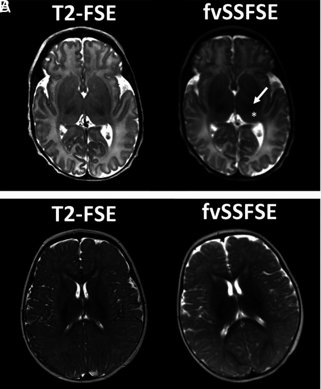
Results: Variable refocusing flip angle single-shot FSE sequences showed more motion artifacts (P < .001). Variable refocusing flip angle single-shot FSE sequences scored higher regarding image sharpness at brain-fluid interfaces (P < .001) and gray-white differentiation (P < .001). Acquisition times for half- and full-Fourier variable refocusing flip angle single-shot FSE were faster than for single-shot FSE (P < .001) with a 53% and 47% reduction, respectively. Intermodality agreement between full-Fourier variable refocusing flip angle single-shot FSE and T2-FSE findings was near-perfect (κ = 0.90, κ = 0.95), with an 8% discordance rate for ground truth lesion detection.

Conclusions: Variable refocusing flip angle single-shot FSE achieved 2× faster scan times than single-shot FSE with improved image sharpness at brain-fluid interfaces and gray-white differentiation. Such improvements are likely attributed to a combination of improved contrast, spatial resolution, SNR, and reduced T2-decay associated with blurring. While variable refocusing flip angle single-shot FSE may be a useful alternative to single-shot FSE and, potentially, T2-FSE when faster scan times are desired, motion artifacts were more common in variable refocusing flip angle single-shot FSE, and, thus, they remain an important consideration before clinical implementation.


Figures

FIG 1.
FIG 1.
TRs of SSFSE and vrfSSFSE. Box-and-whisker plots for each sequence are shown with the middle bar representing the median, the box representing the 25th and 75th percentiles, and the upper and lower bars representing the range. Half-Fourier vrfSSFSE (hvSSFSE) and full-Fourier vrfSSFSE (fvSSFSE) have significantly faster TRs compared with SSFSE (P < . 001). hvSSFSE has a significantly faster TR compared with fvSSFSE (P < . 001). All comparisons of TRs were assessed using the Mann-Whitney test.
FIG 2.
FIG 2.
Image-quality ratings of SSFSE and vrfSSFSE. Box-and-whisker plots for each image-quality parameter (Motion artifact, Sharpness: brain-fluid, and Sharpness: gray-white contrast rated on a 5-point Likert scale) for a given sequence with the middle bar representing the median, the box representing the 25th and 75th percentile, and the upper and lower bars representing the range. A, SSFSE scans scored significantly higher than both hvSSFSE and fvSSFSE scans on motion artifacts (P < . 001). hvSSFSE and fvSSFSE scans did not differ significantly for Motion artifact (P >.5). B, hvSSFSE and fvSSFSE scans were rated significantly better than SSFSE scans for Sharpness: brain-fluid (P < . 001). hvSSFSE and fvSSFSE scans did not differ significantly for Sharpness: brain-fluid (P > .2). C, hvSSFSE and fvSSFSE scans were rated significantly better than SSFSE scans for Sharpness: gray-white contrast (P < . 001). hvSSFSE and fvSSFSE scans did not differ significantly for Sharpness: gray-white contrast (P > .9). All comparisons of image-quality ratings were made using the Wilcoxon matched-pairs signed-rank test.
FIG 3.
FIG 3.
Example of motion-related signal loss. An 8-year-old girl presented for ventricular assessment. Half-Fourier vrfSSFSE (hvSSFSE) and full-Fourier vrfSSFSE (fvSSFSE) images show motion-related signal loss (arrows) on some of the slices that compromised image detail.
FIG 4.
FIG 4.
Example of image sharpness or blurring. A 3-year-old girl presented for ventricular assessment. Half-Fourier vrfSSFSE (hvSSFSE) and full-Fourier vrfSSFSE (fvSSFSE) images (arrows) show improved sharpness or reduced blurring, compared with the corresponding conventional SSFSE, particularly along the cortical margins where the cortical surface is better distinguished against the overlying subarachnoid spaces.
FIG 5.
FIG 5.
Example of magnified views demonstrating differences in tissue contrast. A 2-year-old boy presented for ventricular assessment. Half-Fourier vrfSSFSE (hvSSFSE) and full-Fourier vrfSSFSE (fvSSFSE) images show improved tissue contrast (arrows) that allows improved visualization of the gray-white junction, cerebellar folia detail, as well as posterior hippocampal regions.
FIG 6.
FIG 6.
Normal pediatric infant brain. High tissue contrast that is feasible with full-Fourier vrfSSFSE (fvSSFSE) compared with conventional SSFSE robustly delineates both gray-white differentiation as well as the myelination pattern that nearly resembles that of conventional T2-FSE. Because SSFSE is acquired with long echo trains (during which the signal intensity decays), image blurring occurs. This effect arises from the different signal intensities of each echo, depending on their individual TEs. A, An infant with premature birth history, now near-term in age, presents for screening MR imaging. Note age-appropriate myelination with dark signal in the thalamus (asterisk) and posterior limb of internal capsule (arrow). B, A 6-month-old male infant with spasmus nutans, who presented for screening MR imaging to exclude optic chiasm glioma. In this older infant, myelination has further progressed and is age-appropriate.

References

    1. Iskandar BJ, Sansone JM, Medow J, et al. The use of quick-brain magnetic resonance imaging in the evaluation of shunt-treated hydrocephalus. J Neurosurg 2004;101:147–51 10.3171/ped.2004.101.2.0147 - DOI - PubMed
    1. Ashley WW Jr, McKinstry RC, Leonard JR, et al. Use of rapid-sequence magnetic resonance imaging for evaluation of hydrocephalus in children. J Neurosurg 2005;103:124–30 10.3171/ped.2005.103.2.0124 - DOI - PubMed
    1. Ahn SS, Mantello MT, Jones KM, et al. Rapid MR imaging of the pediatric brain using the fast spin-echo technique. AJNR Am J Neuroradiol 1992;13:1169–77 - PMC - PubMed
    1. Haacke EM, Tkach JA. Fast MR imaging: techniques and clinical applications. AJR Am J Roentgenol 1990;155:951–64 10.2214/ajr.155.5.2120964 - DOI - PubMed
    1. Tekes A, Senglaub SS, Ahn ES, et al. Ultrafast brain MRI can be used for indications beyond shunted hydrocephalus in pediatric patients. AJNR Am J Neuroradiol 2018;39:1515–18 10.3174/ajnr.A5724 - DOI - PMC - PubMed

Publication types