. 2020 Aug;31(8):1746-1760.
doi: 10.1681/ASN.2019101070. Epub 2020 Jun 25.

Central Role for Adipocyte Na,K-ATPase Oxidant Amplification Loop in the Pathogenesis of Experimental Uremic Cardiomyopathy

Affiliations

Central Role for Adipocyte Na,K-ATPase Oxidant Amplification Loop in the Pathogenesis of Experimental Uremic Cardiomyopathy

Komal Sodhi et al. J Am Soc Nephrol. 2020 Aug.

Retraction in

Expression of concern in

Abstract

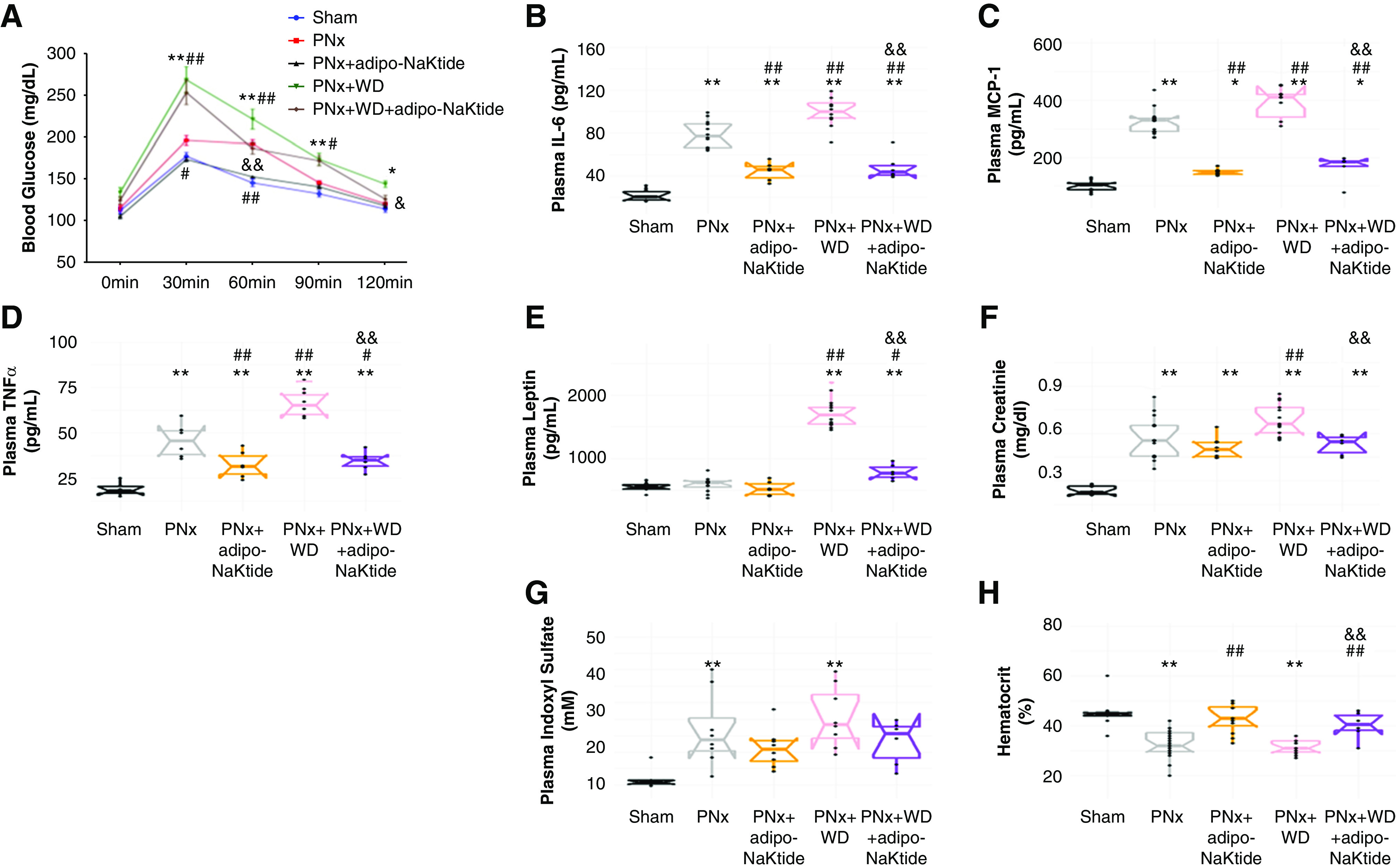
Background: Oxidative stress in adipocyte plays a central role in the pathogenesis of obesity as well as in the associated cardiovascular complications. The putative uremic toxin indoxyl sulfate induces oxidative stress and dramatically alters adipocyte phenotype in vitro. Mice that have undergone partial nephrectomy serve as an experimental model of uremic cardiomyopathy. This study examined the effects on adipocytes of administering a peptide that reduces oxidative stress to the mouse model.

Methods: A lentivirus vector introduced the peptide NaKtide with an adiponectin promoter into the mouse model of experimental uremic cardiomyopathy, intraperitoneally. Then adipocyte-specific expression of the peptide was assessed for mice fed a standard diet compared with mice fed a western diet enriched in fat and fructose.

Results: Partial nephrectomy induced cardiomyopathy and anemia in the mice, introducing oxidant stress and an altered molecular phenotype of adipocytes that increased production of systemic inflammatory cytokines instead of accumulating lipids, within 4 weeks. Consumption of a western diet significantly worsened the adipocyte oxidant stress, but expression of NaKtide in adipocytes completely prevented the worsening. The peptide-carrying lentivirus achieved comparable expression in skeletal muscle, but did not ameliorate the disease phenotype.

Conclusions: Adipocyte-specific expression of NaKtide, introduced with a lentiviral vector, significantly ameliorated adipocyte dysfunction and uremic cardiomyopathy in partially nephrectomized mice. These data suggest that the redox state of adipocytes controls the development of uremic cardiomyopathy in mice subjected to partial nephrectomy. If confirmed in humans, the oxidative state of adipocytes may be a therapeutic target in chronic renal failure.

Keywords: adipocyte; cardiovascular disease; chronic kidney disease; obesity; oxidative stress; uremia.


Figures

Figure 1.
Figure 1.
Immunofluorescence showing colocalization of c-Src (yellow) and NaKtide (red) in PNx+adipo-NaKtide adipose tissue. Images taken with a Leica SP5 TCSII with Coherent Chameleon Multiphoton (MP) Vision II (IR) laser confocal microscope. (A) Adipose images taken with different parameters to select DAPI, cSrc, and NaKtide merged image were obtained. (B) Pixel intensity of the region of interest (ROI) in the different channels was used to calculate the PCC and MOC. Scale bar, 25 µm, ×100 magnification (n=6).
Figure 2.
Figure 2.
Adipo-NaKtide transduction ameliorates visceral adipose oxidative stress and Na,K-ATPase signaling pathways. (A) Thiobarbituric acid–reactive substance, (B) protein carbonylation, (C) pSrc normalized to total Src. mRNA expression of (D) TLR4, (E) TRAF6, (F) PGC1α, and (G) Sirt3. Data are displayed as scatterplots showing data points superimposed on box plots, as described in the Methods. n=5–12/group; *P<0.05 versus sham; **P<0.01 versus sham; #P<0.05 versus PNx; ##P<0.01 versus PNx; &P<0.05 versus PNx+WD; &&P<0.01 versus PNx+WD.
Figure 3.
Figure 3.
Adipo-NaKtide transduction ameliorates visceral adipose inflammatory and apoptotic mRNA. RT-PCR mRNA expression of (A) IL-6, (B) TNF-α, (C) leptin, (D) F4/80, (E) caspase-9, and (F) Bax. (G) NaKtide concentration. Data are displayed as scatterplots showing data points superimposed on box plots, as described in the Methods. n=5–12/group; *P<0.05 versus sham; **P<0.01 versus sham; #P<0.05 versus PNx; ##P<0.01 versus PNx; &P<0.05 versus PNx+WD; &&P<0.01 versus PNx+WD.
Figure 4.
Figure 4.
Adipo-NaKtide transduction improves glucose tolerance and ameliorates circulating inflammatory cytokines, plasma creatinine and indoxyl sulfate, and hematocrit. (A) Glucose tolerance test. Plasma concentrations of (B) IL-6, (C) MCP-1, (D) TNFα, and (E) leptin. Plasma (F) creatinine and (G) indoxyl sulfate concentrations. (H) Hematocrit. n=6–12/group; *P<0.05 versus sham; **P<0.01 versus sham; #P<0.05 versus PNx; ##P<0.01 versus PNx; &P<0.05 versus PNx+WD; &&P<0.01 versus PNx+WD.
Figure 5.
Figure 5.
Adipo-NaKtide transduction improves cardiac phenotype. (A) Heart weight. (B) Representative images and quantification for cardiac fibrosis assessed with Sirius red staining. For this histologic analysis, five spots per section (three sections×five slides per sample) were randomly selected. Images taken with ×20 objective lens; scale bar, 100 µm; n=8–12/group. RT-PCR measurement of mRNA (C) TNFα, (D) IL-6, (E) collagen 1, and (F) Fli-1. (G) Protein carbonylation levels. n=6–12/group; *P<0.05 versus sham; **P<0.01 versus sham; #P<0.05 versus PNx; ##P<0.01 versus PNx; &P<0.05 versus PNx+WD; &&P<0.01 versus PNx+WD.
Figure 6.
Figure 6.
Neither adipo-sNaKtide nor myoD-NaKtide transduction had any effect on cardiac phenotype. (A) Heart weight, (B) plasma creatinine, and (C) hematocrit. Plasma levels of (D) TNFα and (E) MCP1. RT-PCR mRNA expression of (F) IL-6, (G) TNFα, and (H) collagen 1 in heart tissue. n=5–12/group; *P<0.05 versus sham; **P<0.01 versus sham; #P<0.05 versus PNx; ##P<0.01 versus PNx; &P<0.05 versus PNx; &&P<0.01 versus PNx+adipo-NaKtide.

Similar articles

Cited by

References

    1. Bartlett DE, Miller RB, Thiesfeldt S, Lakhani HV, Khanal T, Pratt RD, et al. .: Uremic toxins activates Na/K-ATPase oxidant amplification loop causing phenotypic changes in adipocytes in in vitro models. Int J Mol Sci 19: 2685, 2018 - PMC - PubMed
    1. Sodhi K, Srikanthan K, Goguet-Rubio P, Nichols A, Mallick A, Nawab A, et al. .: pNaKtide attenuates steatohepatitis and Atherosclerosis by blocking Na/K-ATPase/ROS amplification in C57Bl6 and ApoE knockout mice fed a western diet. Sci Rep 7: 193, 2017. - PMC - PubMed
    1. Sodhi K, Maxwell K, Yan Y, Liu J, M.A. Chaudhry MA, Getty M, et al. .: pNaKtide inhibits Na/K-ATPase reactive oxygen species amplification and attenuates adipogenesis. Sci Adv 1: e1500781, 2015. - PMC - PubMed
    1. Furukawa S, Fujita T, Shimabukuro M, Iwaki M, Yamada Y, Nakajima Y, et al. .: Increased oxidative stress in obesity and its impact on metabolic syndrome. J Clin Invest 114: 1752–1761, 2004. - PMC - PubMed
    1. Le Lay S, Simard G, Martinez MC, Andriantsitohaina R: Oxidative stress and metabolic pathologies: From an adipocentric point of view. Oxid Med Cell Longev 2014: 908539, 2014. - PMC - PubMed

Publication types