. 2020 Sep;98(3):222-233.
doi: 10.1124/mol.119.118893. Epub 2020 Jun 25.

Small Molecule Inhibitors Confirm Ubiquitin-Dependent Removal of TOP2-DNA Covalent Complexes

Affiliations

Small Molecule Inhibitors Confirm Ubiquitin-Dependent Removal of TOP2-DNA Covalent Complexes

Rebecca L Swan et al. Mol Pharmacol. 2020 Sep.

Abstract

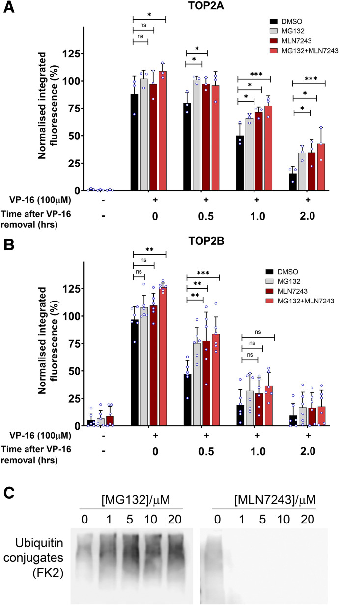
DNA topoisomerase II (TOP2) is required for the unwinding and decatenation of DNA through the induction of an enzyme-linked double-strand break (DSB) in one DNA molecule and passage of another intact DNA duplex through the break. Anticancer drugs targeting TOP2 (TOP2 poisons) prevent religation of the DSB and stabilize a normally transient intermediate of the TOP2 reaction mechanism called the TOP2-DNA covalent complex. Subsequently, TOP2 remains covalently bound to each end of the enzyme-bridged DSB, which cannot be repaired until TOP2 is removed from the DNA. One removal mechanism involves the proteasomal degradation of the TOP2 protein, leading to the liberation of a protein-free DSB. Proteasomal degradation is often regulated by protein ubiquitination, and here we show that inhibition of ubiquitin-activating enzymes reduces the processing of TOP2A- and TOP2B-DNA complexes. Depletion or inhibition of ubiquitin-activating enzymes indicated that ubiquitination was required for the liberation of etoposide-induced protein-free DSBs and is therefore an important layer of regulation in the repair of TOP2 poison-induced DNA damage. TOP2-DNA complexes stabilized by etoposide were shown to be conjugated to ubiquitin, and this was reduced by inhibition or depletion of ubiquitin-activating enzymes. SIGNIFICANCE STATEMENT: There is currently great clinical interest in the ubiquitin-proteasome system and ongoing development of specific inhibitors. The results in this paper show that the therapeutic cytotoxicity of DNA topoisomerase II (TOP2) poisons can be enhanced through combination therapy with ubiquitin-activating enzyme inhibitors or by specific inhibition of the BMI/RING1A ubiquitin ligase, which would lead to increased cellular accumulation or persistence of TOP2-DNA complexes.


Conflict of interest statement

The authors declare that they have no conflict of interest.

Figures

Fig. 1.
Fig. 1.
Effect of inhibiting E1 ubiquitin-activating enzyme on TOP2-DNA complex levels measured using the TARDIS assay. (A and B) K562 cells were treated for 2 hours with etoposide alone or in combination with 10 µM MG132, 10 µM MLN7243, or 10 µM MG132 and 10 µM MLN7243. Cells were incubated for up to a further 2 hours in etoposide-free medium but in the continued presence of DMSO, 10 µM MG132, or 10 µM MLN7243. Levels of TOP2A- and TOP2B-DNA complexes were measured 0, 0.5, 1, and 2 hours after etoposide removal using the TARDIS assay. Median normalized integrated fluorescence values of three independent experiments are shown (circle symbols) along with the mean ± S.D. of the medians. Statistical analyses were performed by two-way ANOVA with comparisons to etoposide-alone treatment using Dunett’s post hoc test. (C) K562 cells incubated with the indicated concentration of MG132 or MLN7243 for 2 hours. Western blots of whole-cell extracts were probed with monoclonal antibody FK2, which recognizes all conjugated ubiquitin (monoubiquinated and polyubiquitinated proteins). ns, not significant.
Fig. 2.
Fig. 2.
The appearance of etoposide-induced DSBs is reduced by chemical inhibition of E1 by inhibitor MLN7243 or by siRNA-mediated depeletion of E1 ubiquitin-activating enzymes. (A) K562 cells were treated with 10 µM etoposide (VP-16) alone or in combination with 10 µM MG132 for up to 4 hours, and protein-free DSBs were detected by γH2AX assay. Median-normalized integrated fluorescence for each replica experiment is indicated by small circle symbols, bars represent the means of the median values ±S.D. (B and C) The γH2AX assay was repeated in K562 cells treated with 10 µM VP-16 alone or in combination with 10 µM MLN7243 for up to 4 hours. Alternatively, cells were treated for 2 hours with etoposide followed by etoposide WO and 2 hours incubation in etoposide-free medium with or without MLN7243 (2 + 2 hours WO). (D) γH2AX levels were quantified after 2 Gy irradiation in the presence and absence of 10 µM MLN7243 and then normalized to a 1-hour postirradiation control. (E and F) γH2AX assay after siRNA silencing of UAE1 and UBA6. All values are normalized to a 1-hour 10 µM etoposide positive control. The medians from independent experiments are shown on each bar. Statistical significance was determined by two-way ANOVA (Bonferroni’s post hoc test). CON, control; DAPI, 4′,6-diamidino-2-phenylindole.
Fig. 3.
Fig. 3.
The effect of siRNA-mediated depletion of UAE1 on the processing of etoposide-induced TOP2-DNA complexes. (A) K562 cells were treated with UAE1 siRNA for 72 hours and then incubated for 2 hours with 100 µM VP-16 or 0.2% DMSO, which was followed by etoposide removal and a further 2 hours incubation in etoposide-free medium. Cells were collected at 0, 0.5, 1, and 2 hours after etoposide removal, and TOP2-DNA complex levels were quantified by TARDIS assay. Statistical comparisons were made by two-way ANOVA with Bonferroni’s post hoc test. (B) siRNA knockdown of UAE1 for 24, 48, 72, and 96 hours in K562 cells was tested by Western blot probing for UAE1.
Fig. 4.
Fig. 4.
MLN7243 inhibits the ubiquitination of TOP2-DNA complexes. (A) K562 cells transfected with control siRNA, UAE1, and UBA6 siRNA or no siRNA were treated with 0.1% DMSO or 10 µM MG132 for 2 hours. Cells without siRNA were also treated with 10 µM MLN7243 to compare levels of ubiquitination activity with UAE1/UBA6 knockdown cells. Total ubiquitination levels were examined by Western blot and probing with clone FK2 antibody, which detects all conjugated ubiquitin. (B) K562 cells were treated with 100 µM etoposide alone or in combination with 10 µM of the UAE inhibitor MLN7243 for 2 hours, and levels of ubiquitinated TOP2-DNA complexes were measured using the TARDIS assay. (C) Control cells (CON siRNA) and double–UAE1/UBA6 siRNA knockdown cells were treated with 100 µM etoposide for 2 hours, and levels of ubiquitinated TOP2-DNA complexes were quantified using the TARDIS assay (n = 3); medians of independent experiments are shown as circles on the bar charts. Statistics was done using two-way ANOVA. Significance comparisons were made by by t test (B) or one-way ANOVA with Tukey’s post hoc test (C).
Fig. 5.
Fig. 5.
BMI1/RING1A is involved in the processing of TOP2-DNA complexes to protein-free DSBs. (A) K562 cells were treated with 100 µM etoposide (VP-16) alone or in combination with 90 µM PRT4165 for 2 hours. Cells were incubated in etoposide-free medium containing DMSO or 90 µM PRT4165 to maintain inhibition of BMI1/RING1A for 0, 0.5, 1, or 2 hours, and levels of TOP2A- and TOP2B-DNA complexes were measured using the TARDIS assay. Averages were normalized to the 2-hour continuous 100 µM etoposide control (0 hours after VP-16 removal), and statistical comparisons were made by two-way ANOVA with Bonferroni’s post hoc test (B) K562 cells were treated with 100 µM etoposide alone or in combination with 90 µM PRT4165 for 2 hours. Cells were collected and processed as per the TARDIS assay, and slides were probed with anti-ubiquitin antibody clone FK2. Averages were normalized to a 2-hour 100 µM etoposide control, and statistical comparisons were made by one-way ANOVA. (C) Cells were treated continuously with 100 µM etoposide alone or in combination with 90 µM PRT4165 for up to 4 hours, and levels of protein-free DSBs were measured using the ɣH2AX assay. Average integrated fluorescence values were normalized to the mean 1-hour 100 µM etoposide treatment value, and statistical comparisons were made by two-way ANOVA with Bonferroni’s post hoc test. Medians of independent experiments are shown as circles on the bar charts. ns, not significant.
Fig. 6.
Fig. 6.
Inhibition of ubiquitin-activating enzyme or BMI1/RING1A sensitizes cells to etoposide. Nalm-6 (A) or Nalm-6TOP2B−/− (B) cells were incubated with increasing concentrations of etoposide (VP-16) alone or in combination with 400 nM MLN7243 for 120 hours. Growth inhibition was determined by XTT assay. IC50 values were determined by plotting dose-response curves; resulting Pf50 values (the fold difference in IC50 values) are shown on the plots. Data plotted are the mean values from at least three separate experiments ±S.E.M. Nalm-6 (C) or Nalm-6TOP2B−/− (D) cells were incubated with increasing concentrations of etoposide (VP-16) alone or in combination with 35 µM PRT4165 for 120 hours. Growth inhibition, IC50, and Pf50 values were determined (A and B). WT, wild type.

Similar articles

Cited by

References

    1. Alchanati I, Teicher C, Cohen G, Shemesh V, Barr HM, Nakache P, Ben-Avraham D, Idelevich A, Angel I, Livnah N, et al. (2009) The E3 ubiquitin-ligase Bmi1/Ring1A controls the proteasomal degradation of Top2alpha cleavage complex - a potentially new drug target. PLoS One 4:e8104. - PMC - PubMed
    1. Aparicio T, Baer R, Gottesman M, Gautier J. (2016) MRN, CtIP, and BRCA1 mediate repair of topoisomerase II-DNA adducts. J Cell Biol 212:399–408. - PMC - PubMed
    1. Atwal M, Swan RL, Rowe C, Lee KC, Lee DC, Armstrong L, Cowell IG, Austin CA. (2019) Intercalating TOP2 poisons attenuate topoisomerase action at higher concentrations. Mol Pharmacol 96:475–484. - PMC - PubMed
    1. Azarova AM, Lyu YL, Lin C-P, Tsai Y-C, Lau JY-N, Wang JC, Liu LF. (2007) Roles of DNA topoisomerase II isozymes in chemotherapy and secondary malignancies. Proc Natl Acad Sci USA 104:11014–11019. - PMC - PubMed
    1. Ban Y, Ho C-W, Lin R-K, Lyu YL, Liu LF. (2013) Activation of a novel ubiquitin-independent proteasome pathway when RNA polymerase II encounters a protein roadblock. Mol Cell Biol 33:4008–4016. - PMC - PubMed

Publication types

MeSH terms

LinkOut - more resources