Randomized Controlled Trial
. 2020 Jun 25;10(1):10344.
doi: 10.1038/s41598-020-67247-4.

Cardioprotective effect of succinate dehydrogenase inhibition in rat hearts and human myocardium with and without diabetes mellitus

Affiliations
Randomized Controlled Trial

Cardioprotective effect of succinate dehydrogenase inhibition in rat hearts and human myocardium with and without diabetes mellitus

Nichlas Riise Jespersen et al. Sci Rep. .

Abstract

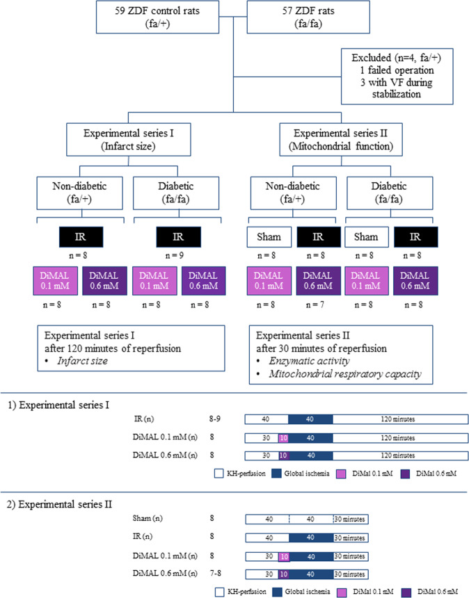
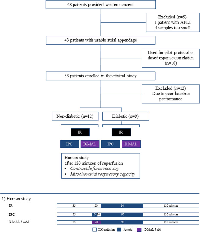
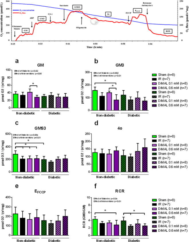
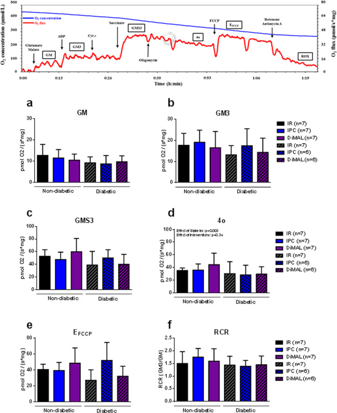
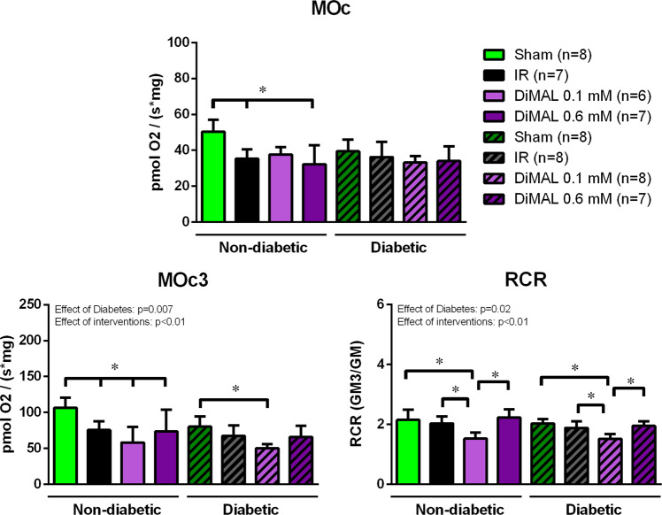
Ischemia reperfusion (IR) injury may be attenuated through succinate dehydrogenase (SDH) inhibition by dimethyl malonate (DiMAL). Whether SDH inhibition yields protection in diabetic individuals and translates into human cardiac tissue remain unknown. In isolated perfused hearts from 24 weeks old male Zucker diabetic fatty (ZDF) and age matched non-diabetic control rats and atrial trabeculae from patients with and without diabetes, we compared infarct size, contractile force recovery and mitochondrial function. The cardioprotective effect of a 10 minutes DiMAL administration prior to global ischemia and ischemic preconditioning (IPC) was evaluated. In non-diabetic hearts exposed to IR, DiMAL 0.1 mM reduced infarct size compared to IR (55 ± 7% vs. 69 ± 6%, p < 0.05). Mitochondrial respiration was reduced by DiMAL 0.6 mM compared to sham and DiMAL 0.1 mM (p < 0.05). In diabetic hearts an increased concentration of DiMAL (0.6 mM) was required for protection compared to IR (64 ± 13% vs. 79 ± 8%, p < 0.05). Mitochondrial function remained unchanged. In trabeculae from humans without diabetes, IPC and DiMAL improved contractile force recovery compared to IR (43 ± 12% and 43 ± 13% vs. 23 ± 13%, p < 0.05) but in patients with diabetes only IPC provided protection compared to IR (51 ± 15% vs. 21 ± 8%, p < 0.05). Neither IPC nor DiMAL modulated mitochondrial respiration in patients. Cardioprotection by SDH inhibition is possible in human tissue, but depends on diabetes status. The narrow therapeutic range and discrepancy in respiration between experimental and human studies may limit clinical translation.


Conflict of interest statement

The authors declare no competing interests.

Figures

Figure 1
Figure 1
Study design of the isolated rat heart study. An overview of the two experimental series of the isolated rat heart study including subgroups and perfusion protocols. IR: Ischemia reperfusion, DiMAL: Dimethyl Malonate, VF: Ventricular fibrillation.
Figure 2
Figure 2
Study design of human study. An overview of the experimental series of the study including subgroups and perfusion protocols. IR: Ischemia reperfusion, IPC: Ischemic preconditioning, DiMAL: Dimethyl Malonate, AFLI: Atrial fibrillation.
Figure 3
Figure 3
Isolated rat heart study – Dose-response relationship and infarct size. Dose-response relationship showing the effect of DiMAL on infarct size (IS) as a percentage of area at risk (AAR) in non-diabetic rat hearts (a) and IS/AAR of different intervention groups in non-diabetic and diabetic ZDF rat hearts (b). IR: Ischemia reperfusion, DiMAL: Dimethyl Malonate. Data are the mean ± SD. *P < 0.05 between indicated groups.
Figure 4
Figure 4
Human study – Dose-response relationship and contractile force recovery. Dose-response relationship between DiMAL and contractile force recovery in non-diabetic human atrial trabeculae (a) and contractile force recovery of non-diabetic and diabetic human atrial trabeculae given as a ratio of contractile force in the reperfusion compared to the pre-ischemic value (b). IR: Ischemia reperfusion, DiMAL: Dimethyl Malonate, IPC: Ischemic preconditioning. Data are mean ± SD. *P < 0.05 between indicated groups. P-values at the top indicate overall effect of diabetes and interventions by two-way ANOVA, respectively.
Figure 5
Figure 5
Isolated rat heart study - Mitochondrial respiratory capacity. Representative graph of mitochondrial respiratory capacity from a Sham heart. Below mitochondrial respiratory capacity. GM, state 2 respiration with glutamate + malate (a); GM3, state 3 respiration with glutamate and malate (b); GMS3, state 3 respiration with glutamate, malate and succinate (c); 4o, state 4 respiration with oligomycin (d); FCCP, non-coupled state by FCCP (e); RCR with complex I-linked substrates (f). Data are mean ± SD. *P < 0.05 between indicated groups. P-values at the top indicate overall effect of diabetes and interventions by two-way ANOVA, respectively.
Figure 6
Figure 6
Human study - Mitochondrial respiratory capacity. Representative graph of mitochondrial respiratory capacity from an IR heart trabecula. Below mitochondrial respiratory capacity. GM, state 2 respiration with glutamate + malate (a); GM3, state 3 respiration with glutamate and malate (b); GMS3, state 3 respiration with glutamate, malate and succinate (c); 4o, state 4 respiration with oligomycin (d); FCCP, non-coupled state by FCCP (e); RCR with complex I-linked substrates (f). Data are mean ± SD. *P < 0.05 between indicated groups. P-values at the top indicate overall effect of diabetes and interventions by two-way ANOVA, respectively.
Figure 7
Figure 7
Isolated rat heart study - Mitochondrial fatty acid respiratory capacity. Summarized data of mitochondrial fatty acid respiratory capacity in each respiratory state. MOc, state 2 respiration with malate and octanoyl-l-carnitine; MOc3, state 3 respiration with malate and octanoyl-l-carnitine; RCR with fatty acids. Data are the mean ± SD. *P < 0.05 between indicated groups. P-values at the top indicate overall effect of diabetes and interventions by two-way ANOVA, respectively.
Figure 8
Figure 8
Isolated rat heart study - Mitochondrial enzyme activity. Enzyme activity of citrate synthase (CS) and β-hydroxyacyl-CoA dehydrogenase (HAD) (a,b). Below protein content of individual electron transport chain complex I-V in non-diabetic (c) and diabetic (d) heart tissue. At the bottom Complex II activity in non-diabetic and diabetic tissue (e) and interstitial succinate concentrations (f). IR: Ischemia reperfusion, DiMAL: Dimethyl malonate. Data are the mean ± SD. *P < 0.05 between indicated groups. P-values at the top indicate overall effect of diabetes and interventions by two-way ANOVA, respectively.

References

    1. Botker HE, Lassen TR, Jespersen NR. Clinical translation of myocardial conditioning. Am. J. Physiol. Heart Circ. Physiol. 2018;314:H1225–H1252. doi: 10.1152/ajpheart.00027.2018. - DOI - PubMed
    1. Di Lisa F, Bernardi P. Mitochondria and ischemia-reperfusion injury of the heart: fixing a hole. Cardiovasc. Res. 2006;70:191–199. doi: 10.1016/j.cardiores.2006.01.016. - DOI - PubMed
    1. Hausenloy, D. J. et al. Ischaemic conditioning and targeting reperfusion injury: a 30 year voyage of discovery. Basic Res. Cardiol. 111, 70-016-0588-8. Epub 2016 Oct 20 (2016). - PMC - PubMed
    1. Heusch G, Botker HE, Przyklenk K, Redington A, Yellon D. Remote ischemic conditioning. J. Am. Coll. Cardiol. 2015;65:177–195. doi: 10.1016/j.jacc.2014.10.031. - DOI - PMC - PubMed
    1. Heusch G. Critical Issues for the Translation of Cardioprotection. Circ. Res. 2017;120:1477–1486. doi: 10.1161/CIRCRESAHA.117.310820. - DOI - PubMed

Publication types

MeSH terms