. 2020 Jun 25;10(1):10355.
doi: 10.1038/s41598-020-67000-x.

Lysophosphatidylcholine acyltransferase 2 (LPCAT2) co-localises with TLR4 and regulates macrophage inflammatory gene expression in response to LPS

Affiliations

Lysophosphatidylcholine acyltransferase 2 (LPCAT2) co-localises with TLR4 and regulates macrophage inflammatory gene expression in response to LPS

W Abate et al. Sci Rep. .

Abstract

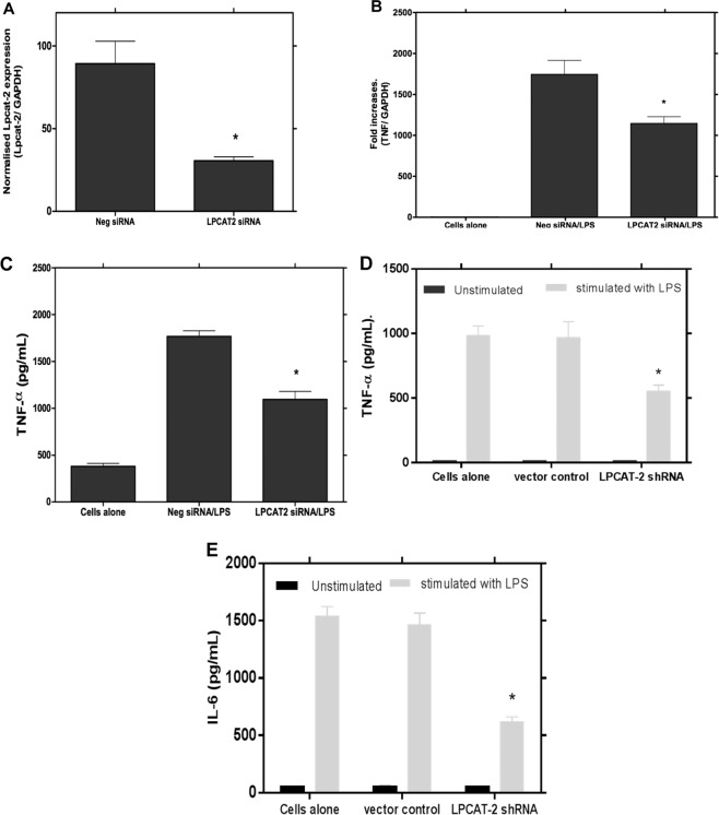
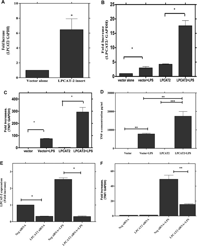
Despite extensive investigations, an effective treatment for sepsis remains elusive and a better understanding of the inflammatory response to infection is required to identify potential new targets for therapy. In this study we have used RNAi technology to show, for the first time, that the inducible lysophosphatidylcholine acyltransferase 2 (LPCAT2) plays a key role in macrophage inflammatory gene expression in response to stimulation with bacterial ligands. Using siRNA- or shRNA-mediated knockdown, we demonstrate that, in contrast to the constitutive LPCAT1, LPCAT2 is required for macrophage cytokine gene expression and release in response to TLR4 and TLR2 ligand stimulation but not for TLR-independent stimuli. In addition, cells transfected to overexpress LPCAT2 exhibited increased expression of inflammatory genes in response to LPS and other bacterial ligands. Furthermore, we have used immunoprecipitation and Western blotting to show that in response to LPS, LPCAT2, but not LPCAT1, rapidly associates with TLR4 and translocates to membrane lipid raft domains. Our data thus suggest a novel mechanism for the regulation of inflammatory gene expression in response to bacterial stimuli and highlight LPCAT2 as a potential therapeutic target for development of anti-inflammatory and anti-sepsis therapies.


Conflict of interest statement

The authors declare no competing interests.

Figures

Figure 1
Figure 1
The level of expression of LPCAT1 and 2 and their induction by LPS or LTA in RAW264.7 cells. The copy number for the LPCATs in RAW264.7 cells were determined as described in the methods section. In resting cells the level of LPCAT1 transcripts was relatively higher than LPCAT2 (A). LPCAT 1 and LPCAT2 mRNA expression in RAW264.7 cells stimulated either LPS (1 µg/mL) (B) or LTA (10 µg/mL) showing the inducibility of LPCAT2 by these ligands (C). n = 3; *p < 0.05 Vs LPCAT1.
Figure 2
Figure 2
Sub-cellular location of LPCAT1 and LPCAT2. Different cellular fractions were isolated and each of the fractions was probed for LPCAT1 or 2 by immunoblotting. Both LPCAT1 and 2 exist in the nuclear- rich fractions (Histone H3 used as a marker) but not in the cytosol (β-tubulin used as cytosolic marker) and their relative abundance does not change following LPS stimulation (A). Isolation of membrane fractions showing that the majority of LPCAT1 and 2 is expressed in the non-raft membrane domain but the level of LPCAT2 is increased in lipid raft domains after LPS stimulation while the level of LPCAT1 remains unchanged (B). Densitometry analysis of the fractions from B are shown for LPCAT2 in (C) and LPCAT1 in (D).
Figure 3
Figure 3
Inhibition of LPCAT2 by siRNA significantly down-regulates LPS-induced inflammatory cytokine gene expression and protein release. Treatment of RAW264.7 cells with LPCAT1 or LPCAT2 siRNA resulted in 81.35 + 6.4 and 80.09 + 1.36 per cent knockdown of LPCAT1 and 2 expression, respectively, compared to the treatment with a control negative siRNA (A). Following siRNA knockdown of LPCAT1 or LPCAT2, cells were stimulated with LPS at 1 µg/mL, for either 4 hours, for cytokine mRNA expression (B–D) or 24 hours for protein release (E,F). *p < 0.05 Vs negative siRNA (B–F).
Figure 4
Figure 4
LPCAT2 silencing reduces LPS-induced TNF-α gene expression and protein release from mouse peritoneal macrophages and a human monocyte cell line. LPCAT2 expression was significantly reduced by incubation of primary mouse peritoneal macrophages with LPCAT2 siRNA (p = 0.0192 vs control negative siRNA) (A). LPCAT2 silenced primary macrophages show a significant reduction in TNF-α gene expression (B) and protein release (C) following stimulation with LPS (100ug/ml). Transduction of the human monocytic cell line, MM6, with shRNA lentiviral particles targeting the LPCAT2 gene resulted in reduced production of TNF-α (D) and IL-6 (E) cytokines following LPS stimulation. *p < 0.05 vs negative siRNA (B,C) vs vector control (D,E).
Figure 5
Figure 5
Overexpression of LPCAT2 gene markedly upregulates LPS-induced TNFα gene expression and protein release. The RAW264 cells were transfected with plasmid carrying the LPCAT2 insert (labelled as LPCAT2), which results in a significant increase in the LPCAT2 gene expression (A). This overexpression is further increased when the cells were stimulated with LPS (B). The overexpression of LPCAT2 significantly increases TNFα gene expression (C) and protein release (D). siRNA silencing significantly reduced LPCAT2 expression in RAW264 cells carrying the LPCAT2 plasmid (E) and significantly inhibited TNFα gene expression in these cells (F). Data represents the mean of four independent experiments (n = 4) ± standard error. *p < 0.05, **p < 0.01, **p < 0.001.
Figure 6
Figure 6
LPCAT2 inhibition had no effect on PMA-induced total reactive oxygen species (ROS) generation while it suppressed LPS-induced ROS production (A). When RAW264.7 cells were stimulated with LPS 30 minutes after they had been transfected with LPCAT2 or control siRNA for 24 hours, LPS-induced activation of p38 was significantly reduced in cells treated with siRNA to LPCAT2 (B). LPS stimulation also induces the physical association between LPCAT2 and TLR4 (C). The physical association of LPCAT2 and TLR4 was demonstrated by immuno-precipitation (IP) after the cells were stimulated with LPS for 15 or 30 minutes. LPCAT2 and TLR4 were detected by immunoblotting following IP using anti-TLR4 and anti-LPCAT2 antibodies respectively (C).

References

    1. Angus DC. The Search for Effective Therapy for Sepsis Back to the Drawing Board? Jama-J Am Med Assoc. 2011;306:2614–2615. doi: 10.1001/jama.2011.1853. - DOI - PubMed
    1. Ward PA. New approaches to the study of sepsis. EMBO molecular medicine. 2012;4:1234–1243. doi: 10.1002/emmm.201201375. - DOI - PMC - PubMed
    1. van der Poll T, van Deventer SJ. Cytokines and anticytokines in the pathogenesis of sepsis. Infectious disease clinics of North America. 1999;13(413–426):ix.. - PubMed
    1. Oberholzer A, Oberholzer C, Moldawer LL. Sepsis syndromes: understanding the role of innate and acquired immunity. Shock. 2001;16:83–96. doi: 10.1097/00024382-200116020-00001. - DOI - PubMed
    1. Moine P, Abraham E. Immunomodulation and sepsis: impact of the pathogen. Shock. 2004;22:297–308. doi: 10.1097/01.shk.0000140663.80530.73. - DOI - PubMed

MeSH terms