. 2020 Jun 25;3(1):330.
doi: 10.1038/s42003-020-1056-4.

Pre-innervated tissue-engineered muscle promotes a pro-regenerative microenvironment following volumetric muscle loss

Affiliations

Pre-innervated tissue-engineered muscle promotes a pro-regenerative microenvironment following volumetric muscle loss

Suradip Das et al. Commun Biol. .

Abstract

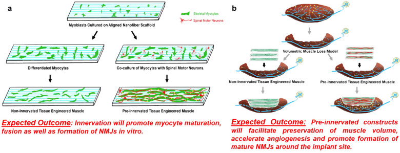
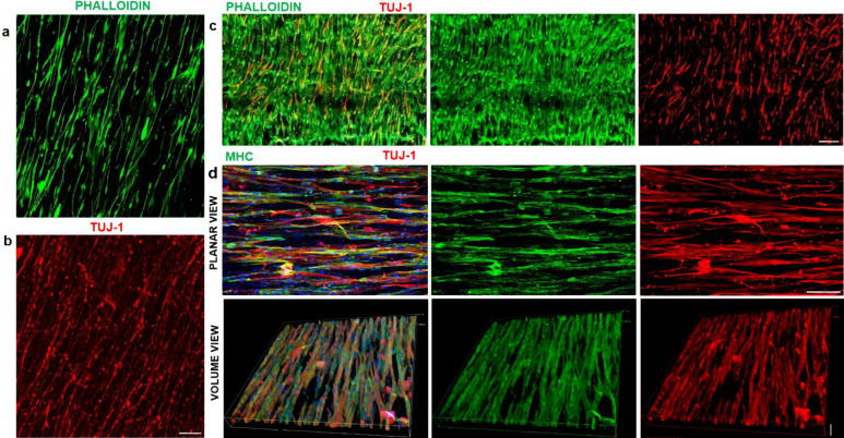
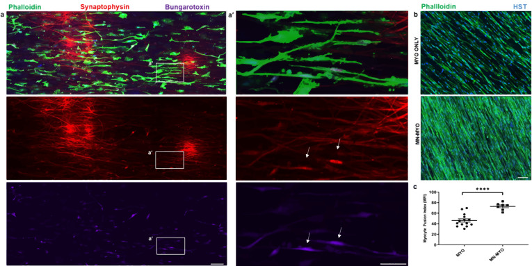
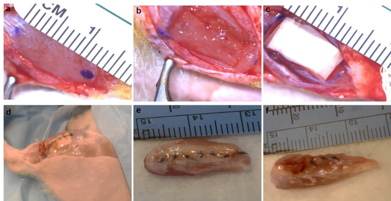
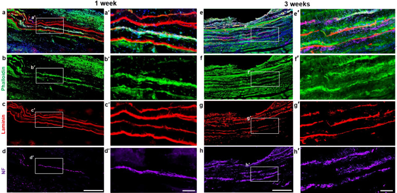
Volumetric muscle loss (VML) is the traumatic or surgical loss of skeletal muscle beyond the inherent regenerative capacity of the body, generally leading to severe functional deficit. Formation of appropriate somato-motor innervations remains one of the biggest challenges for both autologous grafts as well as tissue-engineered muscle constructs. We aim to address this challenge by developing pre-innervated tissue-engineered muscle comprised of long aligned networks of spinal motor neurons and skeletal myocytes on aligned nanofibrous scaffolds. Motor neurons led to enhanced differentiation and maturation of skeletal myocytes in vitro. These pre-innervated tissue-engineered muscle constructs when implanted in a rat VML model significantly increased satellite cell density, neuromuscular junction maintenance, graft revascularization, and muscle volume over three weeks as compared to myocyte-only constructs and nanofiber scaffolds alone. These pro-regenerative effects may enhance functional neuromuscular regeneration following VML, thereby improving the levels of functional recovery following these devastating injuries.


Conflict of interest statement

D.K.C is a co-founder of Axonova Medical, LLC, and INNERVACE, Inc, which are University of Pennsylvania spin-out companies focused on translation of advanced regenerative therapies to treat nervous system disorders. U.S. Provisional Patent App. 62/758,203 (D.K.C., S.D.) has been filed related to the technology of fabricating innervated tissue-engineered constructs. No other author has declared a potential conflict of interest.

Figures

Fig. 1
Fig. 1. Concept of pre-innervated tissue-engineered muscle.
The present study was focused on exploring the role of pre-innervation on myocytes in vitro and host neuromuscular environment in vivo following implantation. a For in vitro studies, our overarching hypothesis was that innervation would augment skeletal myocyte fusion, maturation, and formation of Neuromuscular Junctions (NMJs). b Volumetric Muscle Loss (VML) is defined as frank loss of muscle volume that is accompanied by chronic motor axotomy leading to denervation of the injured area. We used a standardized rat model of VML where >20% of the Tibialis Anterior (TA) muscle volume was resected to create a defect leading to potential damage to intramuscular branches of the host nerve and loss of motor end plates (or NMJs) near the injury area. For in vivo studies, our overarching hypothesis was that implantation of pre-innervated constructs would facilitate preservation of muscle volume, accelerate angiogenesis, enhance Acetylcholine Receptor (AchR) clustering and promote innervation of AchRs (mature NMJs) near the implant site at acute time point.
Fig. 2
Fig. 2. Motor neuron-myocyte culture on aligned nanofibers.
a, b C2C12 Skeletal myocytes and spinal motor neurons were cultured separately as monocultures on aligned PCL nanofiber sheets to optimize media conditions and subsequently stained with Phalloidin and Tuj-1, respectively. Scale bar = 200 μm. c Subsequently, myocytes and motor neurons were cocultured on nanofiber sheets for optimizing coculture media. Scale bar = 200 μm. d In order, to confirm the formation of myotubes and neuromuscular bundles the motor neuron-myocyte cocultures on nanofiber sheets were stained for Myosin Heavy Chain (MHC) and Tuj-1. The neuromuscular bundles were visualized from compressed multiple Z-stack images in planar as well as volumetric view. Scale bar = 100 μm (Planar view), 70 μm (Volume view). The planar and volume view are from the same culture.
Fig. 3
Fig. 3. Innervation of myocytes and effect of motor neurons on myocyte maturation in vitro.
a Rat spinal motor neurons were introduced on a bed of myofibers (Phalloidin) differentiated on aligned nanofiber sheet for 7 days and subsequently cocultured for another 7 days leading to innervation of the skeletal myofibers. Scale bar = 100 µm. a′ Higher magnification view of the area marked by white box reveals structures colabelling for presynaptic marker (Synaptophysin) and Acetylcholine Receptor (AchR) clusters (Bungarotoxin) indicating formation of mature neuromuscular junctions in vitro (indicated by white arrows). Scale bar = 50 µm. b Myocytes exhibited greater fusion when cocultured with motor neurons (MN-MYO) as compared with monoculture (MYO). The cultures were stained with Phalloidin (myocytes) and nuclear marker Hoeschst (HST). Scale bar = 100 µm. c Myocyte Fusion Index (MFI) was calculated from multiple cultures (n ≥ 6), and coculture with motor neurons was found to significantly enhance MFI. For indicated comparison: p ≤ 0.0001 (****). Error bars represent standard error of mean.
Fig. 4
Fig. 4. Bioscaffold implantation in VML model.
a, b Surgical resection of TA muscle to create VML model in rats. c, d Implant of cell-laden nanofiber sheets in muscle defect. Scaffold and overlying fascia secured with sutures. e, f At terminal time points, animals were sacrificed, and TA muscle was excised. Nanofiber sheets were seen in e. Repair Group whereas injury site was recessed in f No Repair group.
Fig. 5
Fig. 5. Pre-innervated bioscaffolds lead to increased recovery of muscle cross-sectional area following VML.
a Representative images demonstrating cross-sectional and longitudinal ultrasound imaging of rat TA muscle. The TA region is demarcated by the yellow line. Multiple nanofiber sheets appeared to be stacked towards the edge of TA in both views as indicated by the white arrows. b, c TA cross-sectional area was measured for MN-MYO (n = 5 for week 1 and week 3), MYO (n = 5 for week 1 and week 3), Sheets (n = 3 for week 1 and week 3) and No Repair (n = 3 for week 1 and week 3) groups by drawing boundaries as indicated by yellow lines. c The recovery of TA CSA was calculated over time across all groups and expressed as percentage of respective contralateral TA CSA. Mean TA CSA percentage as compared to respective contralateral side were as follows – (Week 1) MN-MYO: 91.4; MYO: 87.3; Sheets: 88.7; No Repair: 86.9. (Week 3) MN-MYO: 92.3; MYO: 75.6; Sheets: 74.9; No Repair: 73.2. For indicated comparisons the individual p values were as follows:- MN-MYO vs MYO (Week 3) – p = 0.0033 (**); MN-MYO vs Sheets (Week 3) – p = 0.0089 (**); MN-MYO vs No Repair (Week 3) – p = 0.0037 (**), MYO (Week 1 vs Week 3) – p = 0.0014 (##), SHEETS (Week 1 vs Week 3) – p = 0.0033 (##) and No Repair (Week 1 vs Week 3) – p = 0.0034 (##). Error bars represent standard error of mean.
Fig. 6
Fig. 6. Cellular and morphological evaluation of pre-innervated bioscaffolds following implantation in a VML model.
ah Longitudinal sections near the repair site of animals implanted with nanofibers with motor neurons + myocytes (MN-MYO) at1 week (ad) and 3 weeks (eh) time point. The nanofiber sheets were coated with Laminin prior to culturing cells and hence the stacked sheets were identified based on Laminin stain (red). Scale bar = 500 µm. (a′h′) Magnified view of the corresponding region inside the white box. Thick bundles of myocytes (Phalloidin: green) and motor axons (NF: purple) were observed within the stacked nanofiber sheets both at 1 week and 3 weeks’ time point. Scale bar = 100 µm.
Fig. 7
Fig. 7. Pre-innervated bioscaffolds increase satellite cell density near injury area following VML.
a Muscle satellite cells near the injury area were identified by staining for satellite cell marker – Pax 7 (Purple) across all the groups 1 week and 3 weeks following VML. Scale bar = 100 µm. b Representative image of a higher magnification view of satellite cells. Pax 7+ nuclei (Purple) located on the periphery of Skeletal Muscle Actin + (Red) myofiber and colabelling with pan-nuclear marker Hoeschst (Blue) were identified as satellite cells. Pax-7/HST+ nuclei are indicated by white arrows. Scale bar = 10 µm. c Satellite cell density near the injury area (5 mm2) was counted across MN-MYO (n = 5 for week 1 and week 3), MYO (n = 4 for week 1 and n = 5 for week 3), Sheets (n = 3 for week 1 and week 3) and No Repair (n = 5 for week 1 and n = 3 for week 3) groups. Mean satellite cell density of each group were as follows: (week 1) MN-MYO - 141.4; MYO - 64.0; Sheets – 67.8; No Repair – 66.9; (week 3)) MN-MYO - 224; MYO – 83.2; Sheets – 142.7; No Repair – 94.7. For indicated comparisons the individual p values were as follows: (week 1) MN-MYO vs MYO – p = 0.0029 (**); MN-MYO vs Sheets – p = 0.0081 (**); MN-MYO vs No Repair – p = 0.0024 (**). (Week 3) MN-MYO vs MYO – p < 0.0001 (****); MN-MYO vs Sheets – p = 0.0202 (*); MN-MYO vs No Repair – p = 0.0006 (***). (Week 1 vs Week 3) MN-MYO – p = 0.0034 (##); Sheets – p = 0.034 (#). Error bars represent standard error of mean.
Fig. 8
Fig. 8. Pre-innervated bioscaffolds promote revascularization within and outside the injury area following VML.
a Endothelial cells and microvasculature near the injury area were identified by staining for endothelial cell marker – CD31 (Green) and Smooth Muscle Actin (Red) across all groups 1 week and 3 weeks following VML. The implanted sheets are demarcated by broken white lines. Scale bar = 200 µm. b Representative image of a higher magnification view of endothelial cells and micro-vessels. Structures expressing CD31 (Green) and Smooth Muscle Actin (Red) with a visible lumen and an area > 50 µm2 were defined as micro-vessels. Scale bar = 10 µm. c Microvessel density near the injury area (5 mm2) was counted across MN-MYO (n = 5 for week 1 and week 3), MYO (n = 4 for week 1 and n = 5 for week 3), Sheets (n = 3 for week 1 and week 3) and No Repair (n = 5 for week 1 and n = 3 for week 3) groups. Mean microvessel density of each group were as follows: (Week 1) MN-MYO – 40.9; MYO – 17.7; Sheets – 18.4; No Repair – 25.8. (Week 3) MN-MYO – 116; MYO – 49.6; Sheets – 153.3; No Repair – 112. For indicated comparisons the individual p values were as follows: (Week 1) MN-MYO vs MYO – p = 0.0024 (**); MN-MYO vs Sheets – p = 0.0061 (**); MN-MYO vs No Repair – p = 0.0321 (*). (Week 1 vs Week 3) MN-MYO – p = 0.0049 (##); Sheets – p < 0.0001 (####). No Repair – p = 0.001 (###). Error bars represent standard error of mean. d Microvessel infiltration inside the implanted sheets was evaluated across the repair groups at 3-week time point. Mean number of microvessels inside the sheet region were as follows: MN-MYO – 169.2; MYO – 93; Sheets – 68.33. For indicated comparisons the individual p values were as follows: MN-MYO vs MYO – p = 0.0129 (*); MN-MYO vs Sheets – p = 0.0056 (**). Error bars represent standard error of mean.
Fig. 9
Fig. 9. Acetylcholine receptor (AchR) clusters near injury area following VML.
a AchR clusters near the injury area were identified by staining with Bungarotoxin (Purple) across all groups 1 week and 3 weeks following VML Scale bar =500 µm. b Representative image of a higher magnification view of pretzel shaped AchR clusters (Purple) on the periphery of muscle fibers (Phalloidin-488). Scale bar = 50 µm. c AchR cluster density near the injury area (5 mm2) was counted across MN-MYO (n = 5 for week 1 and week 3), MYO (n = 4 for week 1 and n = 5 for week 3), Sheets (n = 3 for week 1 and week 3) and No Repair (n = 5 for week 1 and n = 3 for week 3) groups. Mean AchR cluster density (per mm2) of each group were as follows: (Week 1) MN-MYO – 5.9; MYO – 2.2; Sheets – 2.3; No Repair – 3.2. (Week 3) MN-MYO – 4.4; MYO – 2.8; Sheets – 1.9; No Repair – 5.6. For indicated comparisons the individual p values were as follows: (Week 1) MN-MYO vs MYO – p < 0.0001 (****); MN-MYO vs Sheets – p = 0.0001 (***); MN-MYO vs No Repair – p = 0.0006 (***). Error bars represent standard error of mean.
Fig. 10
Fig. 10. Pre-innervated bioscaffolds promotes formation of nmjs near injury area following VML.
a NMJs near the injury area were identified by double staining with Bungarotoxin (Purple) and presynaptic marker Synaptophysin (Red) across all groups 1 week and 3 weeks following VML and are indicated by white arrows. Scale bar = 100 µm. b Representative image of a higher magnification view of mature NMJs (indicated by white arrows) comprising of pretzel shaped AchR clusters (Purple) colabelling with presynaptic marker Synaptophysin (Red) located on the periphery of muscle fibers (Phalloidin-488). Scale bar = 10 µm. c Percentage of AchR clusters near the injury area (5 mm2) that were innervated (Synaptophysin+) was counted across MN-MYO (n = 5 for week 1 and week 3), MYO (n = 4 for week 1 and n = 5 for week 3), Sheets (n = 3 for week 1 and week 3) and No Repair (n = 5 for week 1 and n = 3 for week 3) groups to depict maintenance/formation of NMJs in the host muscle. Mean percentage of NMJ of each group were as follows: (Week 1) MN-MYO – 78.9; MYO – 52.9; Sheets –38.7; No Repair – 20.2. (Week 3) MN-MYO – 61.5; MYO – 14.1; Sheets –12.2; No Repair – 18.3. For indicated comparisons the individual p values were as follows: (Week 1) MN-MYO vs MYO – p = 0.0168 (*); MN-MYO vs Sheets – p = 0.0012 (**); MN-MYO vs No Repair – p < 0.0001 (****). (Week 3) MN-MYO vs MYO – p = 0.0004 (***); MN-MYO vs Sheets – p = 0.0011 (**); MN-MYO vs No Repair – p = 0.0031 (**). (Week 1 vs Week 3) MYO – p = 0.0008 (###); Sheets – p = 0.0442 (#). Error bars represent standard error of mean.

References

    1. Kreipke, R. E. & Birren, S. J. Innervating sympathetic neurons regulate heart size and the timing of cardiomyocyte cell cycle withdrawal. J. Physiol. 10.1113/JP270917 (2015). - PMC - PubMed
    1. Andreassen AK. Point:Counterpoint: cardiac denervation does/does not play a major role in exercise limitation after heart transplantation. J. Appl. Physiol. 2008;104:559–560. doi: 10.1152/japplphysiol.00694.2007. - DOI - PubMed
    1. Oberpenning, F., Meng, J., Yoo, J. J. & Atala, A. De novo reconstitution of a functional mammalian urinary bladder by tissue engineering. Nat. Biotechnol. 10.1038/6146 (1999). - PubMed
    1. Morimoto, Y., Kato-Negishi, M., Onoe, H. & Takeuchi, S. Three-dimensional neuron-muscle constructs with neuromuscular junctions. Biomaterials10.1016/j.biomaterials.2013.08.062 (2013). - PubMed
    1. Guo, X., Gonzalez, M., Stancescu, M., Vandenburgh, H. H. & Hickman, J. J. Neuromuscular junction formation between human stem cell-derived motoneurons and human skeletal muscle in a defined system. Biomaterials10.1016/j.biomaterials.2011.09.014(2011). - PMC - PubMed

Publication types