. 2020 Jun 25;10(1):10343.
doi: 10.1038/s41598-020-67143-x.

TET1 Deficiency Impairs Morphogen-free Differentiation of Human Embryonic Stem Cells to Neuroectoderm

Affiliations

TET1 Deficiency Impairs Morphogen-free Differentiation of Human Embryonic Stem Cells to Neuroectoderm

Hanqin Li et al. Sci Rep. .

Abstract

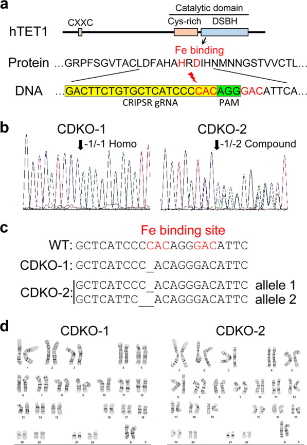
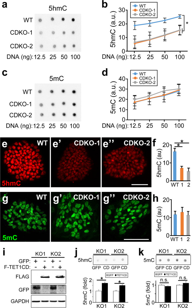
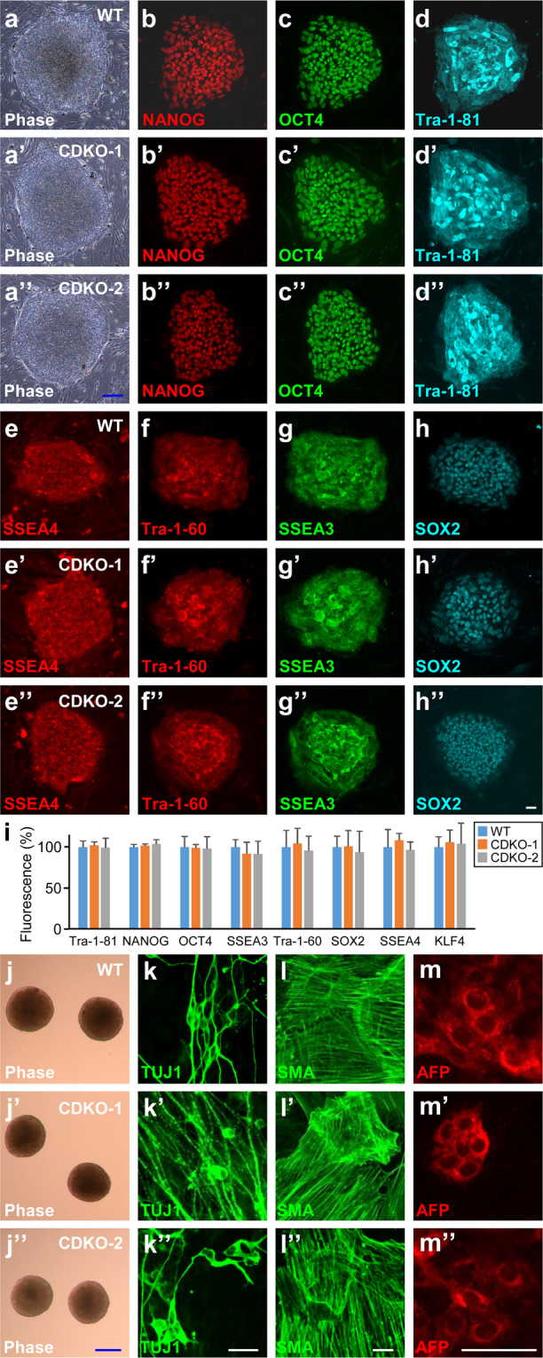
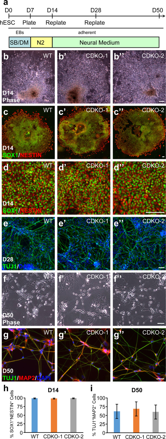
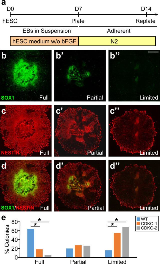
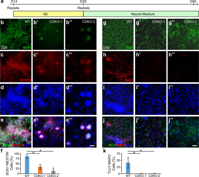
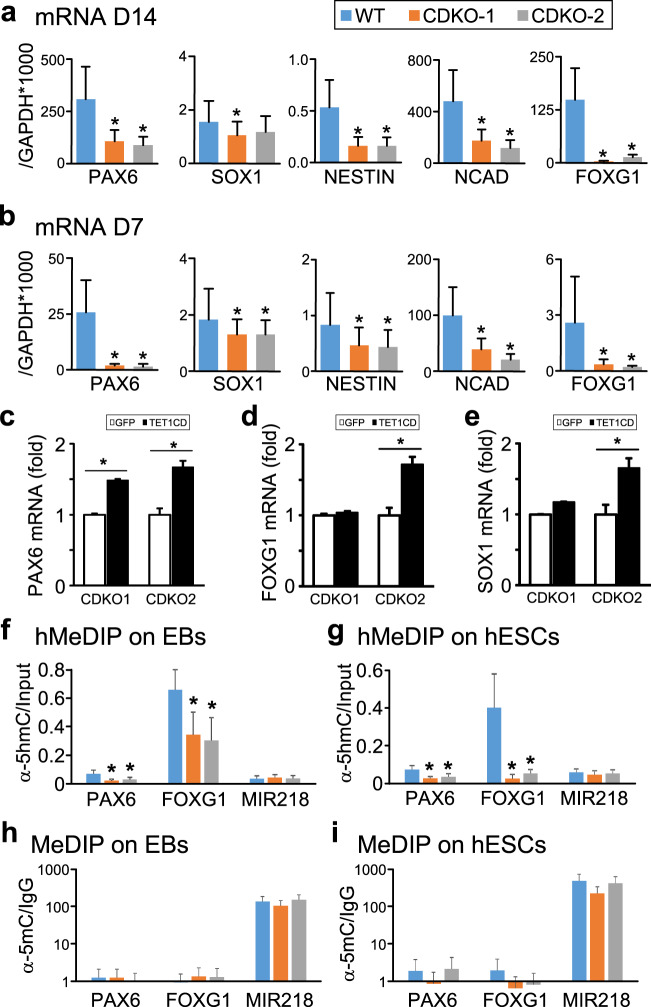
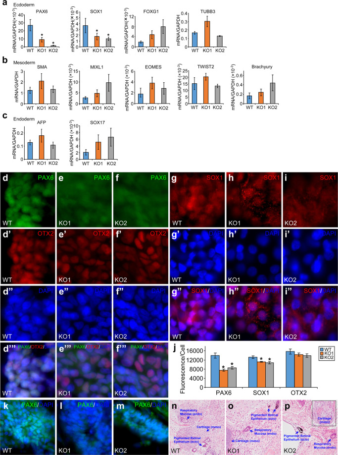
The TET family of 5-methylcytosine (5mC) dioxygenases plays critical roles in development by modifying DNA methylation. Using CRISPR, we inactivated the TET1 gene in H9 human embryonic stem cells (hESCs). Mutant H9 hESCs remained pluripotent, even though the level of hydroxymethylcytosine (5hmC) decreased to 30% of that in wild-type cells. Neural differentiation induced by dual SMAD inhibitors was not significantly affected by loss of TET1 activity. However, in a morphogen-free condition, TET1 deficiency significantly reduced the generation of NESTIN+SOX1+ neuroectoderm cells from 70% in wild-type cells to 20% in mutant cells. This was accompanied by a 20-fold reduction in the expression level of PAX6 and a significant decrease in the amount of 5hmC on the PAX6 promoter. Overexpression of the TET1 catalytic domain in TET1-deficient hESCs significantly increased 5hmC levels and elevated PAX6 expression during differentiation. Consistent with these in vitro data, PAX6 expression was significantly decreased in teratomas formed by TET1-deficient hESCs. However, TET1 deficiency did not prevent the formation of neural tube-like structures in teratomas. Our results suggest that TET1 deficiency impairs the intrinsic ability of hESCs to differentiate to neuroectoderm, presumably by decreasing the expression of PAX6, a key regulator in the development of human neuroectoderm.


Conflict of interest statement

The authors declare no competing interests.

Figures

Figure 1
Figure 1
CRISPR-mediated mutations of catalytic domain of TET1 in H9 hESCs. (a) Double strand break was introduced by CRISPR/Cas9 before the first iron binding site to induce non-homologous end joining in TET1 gene. (b) Sequencing traces of PCR products amplified from genomic DNA of two hESC clones (CDKO-1 and CDKO-2), showing frame shift mutations in both alleles in each clone. (c) Sequencing of cloned PCR products showing homozygous 1 bp deletion in CDKO-1 clone and compound heterozygous deletions of 1 bp and 2 bp in CDKO-2 clone. The frameshift mutations destroy iron-binding sites and downstream catalytic domain. (d) Normal karyotype for the two TET1 mutant clones.
Figure 2
Figure 2
Reduced 5hmC level in TET1-deficient hESCs. (a,b) Dot blot (a) and quantification (b) of 5hmC in WT and TET1-deficient hESCs. *p < 0.001, paired Student’s t-test, n = 3. (c,d) Dot blot (c) and quantification of 5mC in WT and TET1-deficient hESCs. Full images of the dot blots without any imaging processing are displayed in (a) and (c). (e,f) 5hmc staining (e-e”) and quantification (f) in WT (e) and TET1-deficient (e’ and e”) hESCs. *p < 0.001, Student’s t-test, n = 5. (g,h) 5mC staining (g-g”) and quantification (h) in WT (g) and TET1-deficient (g’ g”) hESCs. Scale bar, 100 µm. (i,-k) The two TET1 mutant hESC lines (KO1 and KO2) were infected with lentivirus expressing GFP or FLAG-tagged TET1 catalytic domain (F-TET1CD). Total cell lysates were blotted with the indicated antibodies (i). Genomic DNA isolated from these cells were dot-blotted with antibodies against 5hmC (j) or 5mC (k). *p < 0.05, paired Student’s t-test, n = 4.
Figure 3
Figure 3
Pluripotency maintained in TET1-deficient hESCs. (a–i) Phase contrast images (a-a”) and immunostaining of indicated pluripotency markers (b-h”) in wild-type (a–h) or TET1-deficient (a’-h”) H9 hESCs. Fluorescence intensity was quantified from at least five independent colonies for each condition and normalized against the values in WT (i). (j–m”) Wild-type (j–m) or TET1-deficient (j’-m) H9 hESCs were differentiated spontaneously in serum-containing medium through embryoid bodies (j–j”) to cells of all three germ layers, as indicated by immunostaining for the ectoderm marker TUJ1 (k-k”), the mesoderm marker SMA (l-l”) and the endoderm marker AFP (m-m”). Blue bars, 100 µm; white bars, 25 µm.
Figure 4
Figure 4
Normal neural differentiation of TET1-deficient hESCs under dual SMAD inhibition. (a) Diagram of neural differentiation protocol using the two SMAD inhibitors SB431542 (SB) and dorsomorphin (DM). EB, Embryoid Body. N2, N2 supplements. (b-d”) Phase contrast images (b-b) and immunostaining for neural lineage markers SOX1 and NESTIN (c-d”) of EB-derived colonies at D14 from the indicated hESC lines. Representative colonies stained in (c-c”) were magnified in (d-d”) accordingly. (e-e”) Immunostaining of the pan-neural marker TUJ1 at D28. (f-g”) Phase contrast images (f-f”) and immunostaining of TUJ1 and MAP2 (g-g”) of mature neurons at D50. (h) Quantification of SOX1+NESTIN+ neuroectoderm cells at D14 from 3 independent experiments. (i) Quantification of TUJ1+MAP2+ mature neurons at D50 from 3 independent experiments. Bars, 100 µm.
Figure 5
Figure 5
Impairment at the early stage of neural differentiation of TET1-deficient hESCs under the morphogen-free condition. (a) Protocol for the differentiation of embryoid bodies (EB) to early neural lineage under morphogen-free condition from day 0 to day 14. N2, N2 supplements. (b–d”) Three types of neuroepithelial colonies were classified by the percentage of SOX1+ NESTIN+ cells among all cells within individual colony, representing the completeness of neural lineage commitment. Full, colonies with a SOX1+NESTIN+ percentage >70% (b–d for separate channels and merged image); Partial, colonies with a SOX1+NESTIN+ percentage between 20–70% (b’-d’); Limited, colonies with a SOX1+NESTIN+ percentage <20% (b”-d”). Bar, 500 µm. (e) Percentage of each type of colonies formed at D14 by WT and the two TET1-deficient lines of H9 hESCs. *p < 0.001; Fisher’s exact test, n = 40 colonies for each cell line.
Figure 6
Figure 6
TET1-deficient hESCs cannot be differentiated to neurons in the morphogen-free condition. (a) Protocol for the differentiation of neuroepithelial cells to neurons in morphogen-free condition. N2, N2 supplements. (b–f) At day 28 of differentiation, neuroectoderm cells generated from wild-type (b–e) or TET1-deficient (b’-e”) H9 hESCs were costained for SOX1 (b–b”), NESTIN (c–c”), and DAPI (d–d”). Merged images (e–e”) were used to quantify the percentage of SOX1 and NESTIN double positive cells among all DAPI+ cells (f). *p < 0.001, Student’s t-test, n = 3. (g–k) At day 50, cells differentiated from wild-type or TET1-deficient H9 hESCs were co-stained for the pan-neural marker TUJ1 (g–g”), mature neuronal marker MAP2 (h–h”), and DAPI (i–i”). Merged images (j–j”) were used to quantify the percentage of MAP2 and TUJ1 double positive neuron among all DAPI+ cells (k). *p < 0.001. Student’s t-test, n = 3. Bars, 100 µm.
Figure 7
Figure 7
Impaired neural differentiation of TET1-deficient hESCs is accompanied by decreased 5hmC content in PAX6 promoter. (a,b) qRT-PCR measurement of the expression levels of the indicated neural lineage markers in cells differentiated from wild-type (WT) or TET1-deficient (CDKO-1 and CDKO-2) H9 hESCs under the morphogen-free condition at day 14 (a) and day 7 (b). *p < 0.05, paired Student’s t-test, n = 3. (c–e) The two TET1 mutant hESC lines (CDKO1 and CDKO2) infected with lentiviruses expressing GFP or FLAG-TET1CD were differentiated in the morphogen-free condition. Embryoid bodies at day 10 of differentiation were analyzed by qRT-PCR for the expression levels the neuroecdoderm genes PAX6 (c), FOXG1 (d) and SOX1 (e). *p < 0.05, paired Student’s t-test, n = 6. (f,g) hMeDIP measurement of 5hmC content at the promoter region of PAX6, FOXG1 or miR218, a gene unrelated to early neural differentiation, in EBs formed under morphogen-free condition (f) or in undifferentiated hESCs (g). *p < 0.05, Student’s t-test, n = 3. (h,i) MeDIP measurement of 5mC content at the promoter of PAX6, FOXG1 or miR218 in EBs formed under morphogen-free condition (h) or in undifferentiated hESCs (i).
Figure 8
Figure 8
Reduced expression of PAX6 and SOX1 in teratomas formed by TET1-deficient hESCs. (ac) Quantitative RT-PCR measurement of the expression levels of marker genes for ectoderm (a), mesoderm (b), or endoderm (c) in total RNA isolated from teratomas formed by TET1-deficient (KO1 and KO2) or wild-type (WT) H9 hESCs. *p < 0.05, vs. WT, n = 8–14. No significant difference between KO1 and KO2. (dj) Cryostat sections from these teratomas were costained for PAX6, OTX2 and DNA (d–f’”) or for SOX1 and DNA (g–i”). Average fluorescence intensities per cell for PAX6, SOX1 and OTX2 in these sections were quantified (j). *p < 0.05, vs. WT, n = 12. No significant difference between KO1 and KO2. (k–m) PAX6+ neural tube-like structures in cryostat sections from these teratomas. (n–p) H&E staining of paraffin sections of these teratomas. ecto, ectoderm; meso, mesoderm; endo, endoderm.

Similar articles

Cited by

References

    1. Thomson JA, et al. Embryonic stem cell lines derived from human blastocysts. Science (New York, N.Y.) 1998;282:1145–1147. doi: 10.1126/science.282.5391.1145. - DOI - PubMed
    1. Fox IJ, et al. Stem cell therapy. Use of differentiated pluripotent stem cells as replacement therapy for treating disease. Science (New York, N.Y.) 2014;345:1247391. doi: 10.1126/science.1247391. - DOI - PMC - PubMed
    1. Meissner A. Epigenetic modifications in pluripotent and differentiated cells. Nature biotechnology. 2010;28:1079–1088. doi: 10.1038/nbt.1684. - DOI - PubMed
    1. Boland MJ, Nazor KL, Loring JF. Epigenetic regulation of pluripotency and differentiation. Circulation research. 2014;115:311–324. doi: 10.1161/circresaha.115.301517. - DOI - PMC - PubMed
    1. Koh KP, Rao A. DNA methylation and methylcytosine oxidation in cell fate decisions. Current opinion in cell biology. 2013;25:152–161. doi: 10.1016/j.ceb.2013.02.014. - DOI - PMC - PubMed

Publication types

MeSH terms