. 2020 Jun 25;10(1):10356.
doi: 10.1038/s41598-020-66602-9.

Amylin and beta amyloid proteins interact to form amorphous heterocomplexes with enhanced toxicity in neuronal cells

Affiliations

Amylin and beta amyloid proteins interact to form amorphous heterocomplexes with enhanced toxicity in neuronal cells

Prashant Bharadwaj et al. Sci Rep. .

Abstract

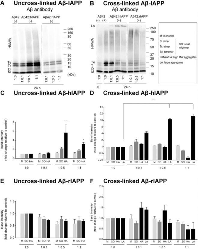
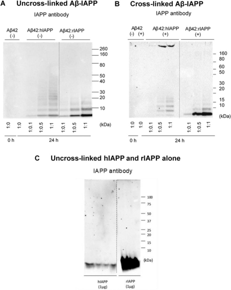
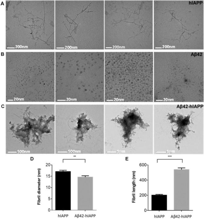
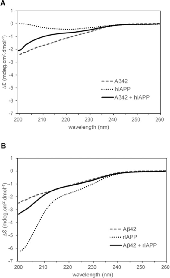
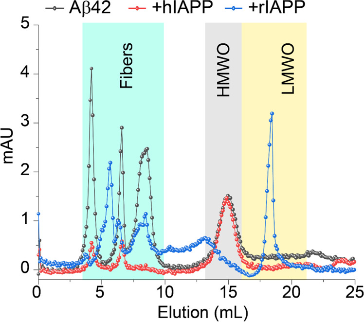
Human pancreatic islet amyloid polypeptide (hIAPP) and beta amyloid (Aβ) can accumulate in Type 2 diabetes (T2D) and Alzheimer's disease (AD) brains and evidence suggests that interaction between the two amyloidogenic proteins can lead to the formation of heterocomplex aggregates. However, the structure and consequences of the formation of these complexes remains to be determined. The main objective of this study was to characterise the different types and morphology of Aβ-hIAPP heterocomplexes and determine if formation of such complexes exacerbate neurotoxicity. We demonstrate that hIAPP promotes Aβ oligomerization and formation of small oligomer and large aggregate heterocomplexes. Co-oligomerized Aβ42-hIAPP mixtures displayed distinct amorphous structures and a 3-fold increase in neuronal cell death as compared to Aβ and hIAPP alone. However, in contrast to hIAPP, non-amyloidogenic rat amylin (rIAPP) reduced oligomer Aβ-mediated neuronal cell death. rIAPP exhibited reductions in Aβ induced neuronal cell death that was independent of its ability to interact with Aβ and form heterocomplexes; suggesting mediation by other pathways. Our findings reveal distinct effects of IAPP peptides in modulating Aβ aggregation and toxicity and provide new insight into the potential pathogenic effects of Aβ-IAPP hetero-oligomerization and development of IAPP based therapies for AD and T2D.


Conflict of interest statement

The authors declare no competing interests.

Figures

Figure 1
Figure 1
Aggregation kinetics of Aβ42-IAPP mixtures. (A) Individual aggregation kinetics of disaggregated Aβ42, hIAPP and rIAPP (20 µM) alongside TBS control assessed by Thioflavin-T (ThT) fluorescence over a 26 h period (mean ± SEM, n = 3, p < 0.001). (B) Aggregation kinetics of Aβ42 (20 µM) co-incubated with different concentrations (1:0.1, 1:0.5, 1:1) of hIAPP and rIAPP assessed by ThT fluorescence over 26 h. ThT fluorescence is represented as arbitrary units (AU; mean ± SEM, n = 3, p < 0.001). With increasing concentrations of hIAPP, Aβ42-hIAPP mixtures demonstrate a dose-dependent increase in ThT fluorescence. Aβ42-rIAPP mixtures demonstrate low ThT fluorescence at all concentrations compared to Aβ42, but this effect was not significant.
Figure 2
Figure 2
Oligomerization and size distribution of uncross-linked and photo cross-linked Aβ42-IAPP mixtures. Western immunoblotting analysis of (A) uncross-linked (−) and (B) cross-linked (+) Aβ42-IAPP at 24 h time-point assessed using Aβ antibody WO2. Densiometric analysis of (C) Aβ42-hIAPP (−), (D) Aβ42-hIAPP (+) (E) Aβ42-rIAPP (−) and (F) Aβ42-rIAPP (+) mixtures at 24 h timepoint to determine levels of monomers (4.5 kDa), small oligomers (9–17 kDa), HMW aggregates (40–160 kDa) and large aggregates (>250 kDa). Marked increase in the formation of small oligomers (2-fold) and HMW aggregates (6-fold) were observed in Aβ42:hIAPP mixtures of 1:0.5 and 1:1 ratios, while Aβ42-rIAPP mixtures did not demonstrate any significant changes as compared to Aβ42 alone. Band intensities represented as fold-change relative to control (mean ± SEM (n = 5), *p < 0.005, ***p < 0.001). Cross-linked samples of Aβ42 incubated with higher concentrations of hIAPP (1:0.5, 1:1) demonstrated a significant 20-fold increase in large aggregates (>250 kDa), combined with a decrease in the levels of oligomers (B,D). An increase in HMW aggregates in Aβ42:rIAPP (1:0.1) and to a lesser extent Aβ42:rIAPP (1:05) was observed, but was not significant (B). Dotted lines indicate spliced bands from the same gel in which blank lanes were removed.
Figure 3
Figure 3
Size distribution of photo cross-linked Aβ42-IAPP heterocomplexes. Western immunoblotting analysis of (A) uncross-linked (−) and (B) cross-linked (+) Aβ42-IAPP combinations at 24 h timepoint assessed using an IAPP antibody (T-4157). (C) uncross-linked hIAPP and rIAPP assessed using IAPP antibody (T-4157). Aβ42 incubated with hIAPP at ratios 1:0.5 and 1:1 showed prominent small oligomers (dimer, trimer and tetramer) and large aggregates (>250 kDa), which were otherwise absent in hIAPP alone (compare A–C). The IAPP antibody detected formation of oligomers in Aβ-rIAPP mixtures, which were absent in rIAPP only, suggesting Aβ42 and rIAPP may be interacting to form heterocomplexes as well. Dotted lines indicate spliced bands from the same gel in which blank lanes were removed.
Figure 4
Figure 4
Electron micrographs of hIAPP, Aβ42 and Aβ42-hIAPP morphology. Electron micrographs demonstrating (A) fibril-like hIAPP (17 ± 0.5 nm in width, 202 ± 9.9 nm in length, n = 77) and spherical (B) Aβ42 oligomers (6 ± 0.2 nm in diameter, n = 207). (C) Aβ42-hIAPP formed large amorphous aggregates with distinct morphologies. Analysis of fibril (D) length and (E) diameter demonstrated that Aβ42-hIAPP (14 ± 0.6 nm in diameter, 539 ± 22.7 nm in length, n = 100) was significantly different from hIAPP (mean ± SEM, **p < 0.005, ***p < 0.001). Scale bar (A) 200 nm, (B) 20 nm, (C) 500 nm/1 µm. Aβ42-hIAPP mixtures are large amorphous structures that are distinctly different from either spherical Aβ42 oligomers or fibril-like hIAPP.
Figure 5
Figure 5
CD spectra Aβ42, IAPP and Aβ42-IAPP combinations between 200–260 nm. (A) CD spectra of Aβ42, hIAPP and Aβ42-hIAPP (1:1) mixtures. hIAPP had spectra characteristic for samples containing predominantly β-sheet, while Aβ42 and Aβ42-hIAPP contained predominantly random coil. (B) CD spectra obtained for Aβ42, rIAPP and Aβ42-rIAPP (1:1) mixtures demonstrated predominantly random coil. Each spectrum represents three biological replicates and nine technical replicates.
Figure 6
Figure 6
Monitoring the aggregation kinetics of Aβ42-IAPP combinations by 1H NMR. (AC) Time-lapse 1H NMR spectra of 25 µM Aβ42 co-incubated with 25 µM hIAPP or rIAPP. NMR samples were prepared using 10 mM NaPi buffer, pH = 7.4 containing 10% D2O and spectra were recorded on a 500 MHz spectrometer at 25 °C. (DF) Decay of 1H NMR signal intensities calculated from the spectra shown in (AC) as a function of time as indicated by colors. The integrated total signal intensities of the selected regions highlighted in (AC) were analyzed using MestReNova.
Figure 7
Figure 7
Size profiling of Aβ42-IAPP combinations using SEC. Size-profile analysis using SEC of 50 µM Aβ42 co-incubated with 50 µM hIAPP or rIAPP as indicated in colors. The SEC samples were prepared using 10 mM NaPi buffer, pH = 7.4 and incubated for ~ 24 h under continuous agitation prior injection onto the column. The HMWO and LMWO refer to the high-molecular and low-molecular weight oligomers, respectively.
Figure 8
Figure 8
Toxicity of co-oligomerized Aβ42-IAPP mixtures in neuronal cells. Toxicity of Aβ42 and IAPP peptide mixtures in human neuroblastomas (SH-SY5Y) was assessed by MTS (soluble tetrazolium) assay. Cells were treated with (A) increasing concentrations (10, 20 and 30 µM) of Aβ42, hIAPP and Aβ42 + hIAPP (1:1) mixtures or (B) Aβ42 co-oligomerized with increasing concentration ratios of IAPP peptides (1:0.1, 1:0.5 and 1:1). Aβ42, hIAPP and Aβ42 + hIAPP treatment showed dose dependent cell death compared to rIAPP and Aβ42 + rIAPP, as indicated by the reduced ability of neurons to metabolize MTS (*p < 0.05, **p < 0.01). At higher concentrations (20 and 30 µM), Aβ42 + hIAPP showed significantly higher cell death as compared to Aβ42 and hIAPP only (##p < 0.01). In contrast to hIAPP, rIAPP rescued cells from Aβ42 induced toxicity. No significant loss of cell viability was observed with different concentrations of Aβ42-rIAPP treatment. At higher concentration ratios of hIAPP (1:0.5 and 1:1), Aβ42-hIAPP mixtures showed a 3–4 fold increase, whereas rIAPP showed a dose dependent decrease in cell death with increase concentrations of rIAPP in Aβ42-rIAPP mixtures.
Figure 9
Figure 9
Toxicity of co-oligomerized and individually oligomerized Aβ42-IAPP mixtures. Toxicity of co-oligomerized and independently oligomerized Aβ42 and IAPP peptides were assessed in human neuroblastomas (SH-SY5Y) was assessed by MTS (soluble tetrazolium) assay. Co-oligomerized Aβ42-hIAPP showed 3–4 fold increase in cell death as compared to Aβ42 only (**p < 0.01). However, when individually oligomerized, Aβ42-hIAPP treatments showed less cell death as compared to Aβ42 only (##pp < 0.01). Notably, rIAPP rescued Aβ42 induced toxicity in a dose-dependent manner in both pre-incubated and non-pre-incubated conditions tested (**p < 0.01).

References

    1. Ninomiya T. Diabetes mellitus and dementia. Curr Diabetes Rep. 2014;14:1–9. - PubMed
    1. Ott A, et al. Association between diabetes mellitus and dementia: the Rotterdam study. Diebetologia. 1996;39:1392–1397. - PubMed
    1. Lutz, T. A. & Meyer, U. Amylin at the interface between metabolic and neurodegenerative disorders. Front Neurosci9 (2015). - PMC - PubMed
    1. Verdile G, Fuller SJ, Martins R. The role of Type 2 diabetes in neurodegeneration. Neurobiol Dis. 2015;84:22–38. - PubMed
    1. Bharadwaj P, et al. The link between Type 2 diabetes and neurodegeneration: roles for amyloid-β, amylin, and tau proteins. J Alzheimer Dis. 2017;59:421–432. - PubMed

Publication types

MeSH terms