. 2020 Jun 10:2020:4359305.
doi: 10.1155/2020/4359305. eCollection 2020.

Amelioration of Coagulation Disorders and Inflammation by Hydrogen-Rich Solution Reduces Intestinal Ischemia/Reperfusion Injury in Rats through NF- κ B/NLRP3 Pathway

Affiliations

Amelioration of Coagulation Disorders and Inflammation by Hydrogen-Rich Solution Reduces Intestinal Ischemia/Reperfusion Injury in Rats through NF- κ B/NLRP3 Pathway

Ling Yang et al. Mediators Inflamm. .

Abstract

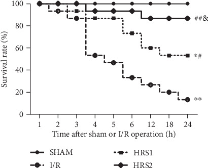
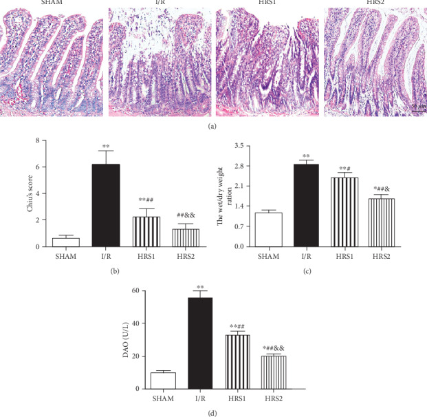
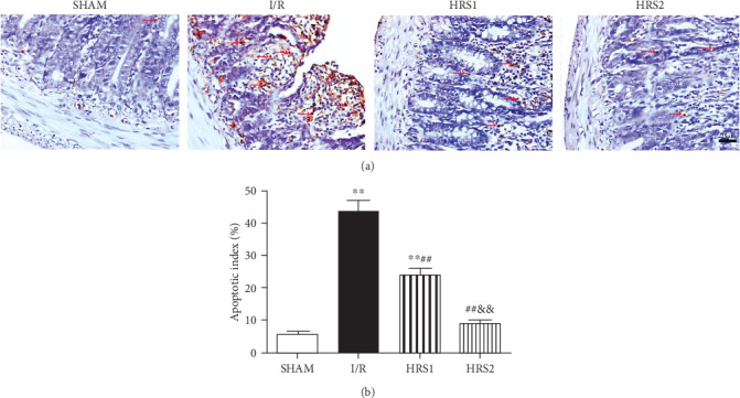
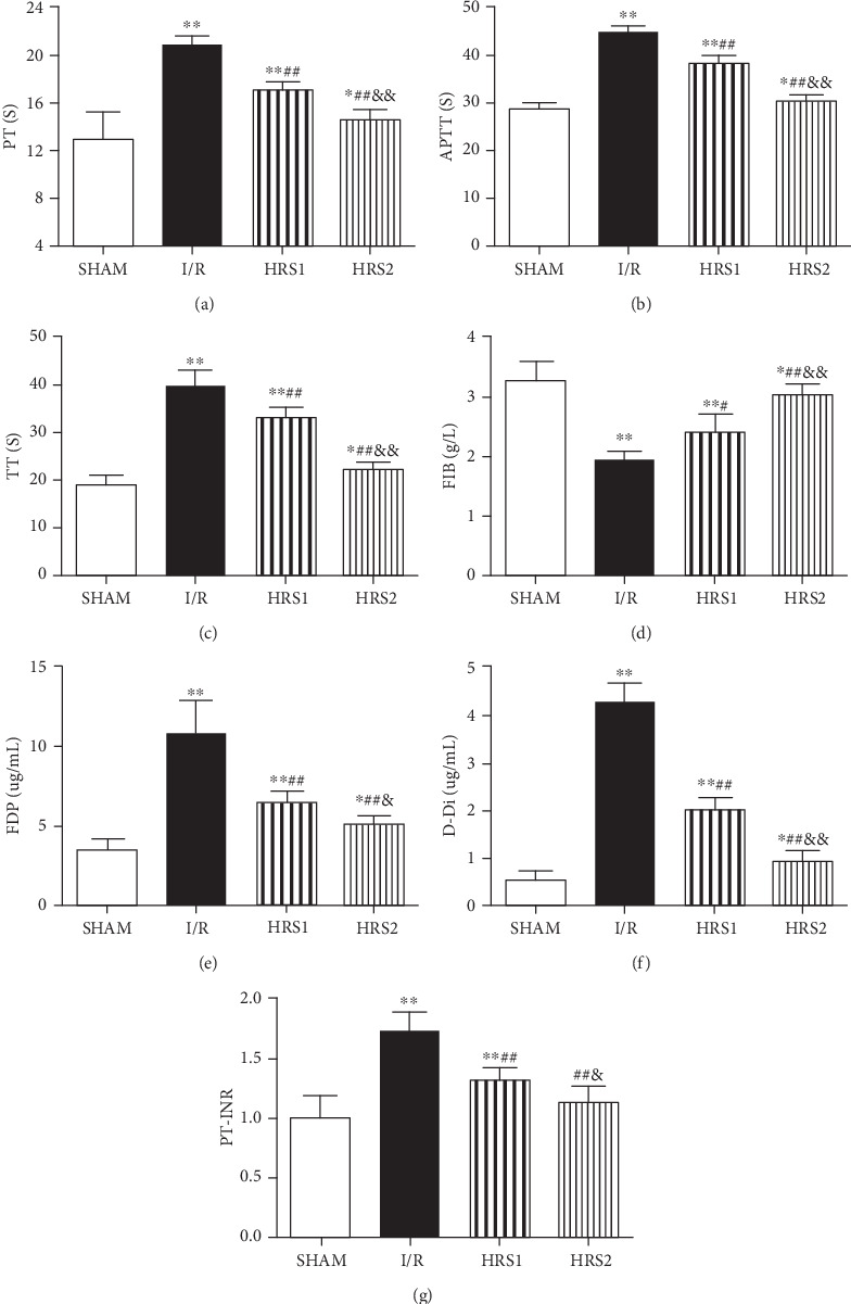
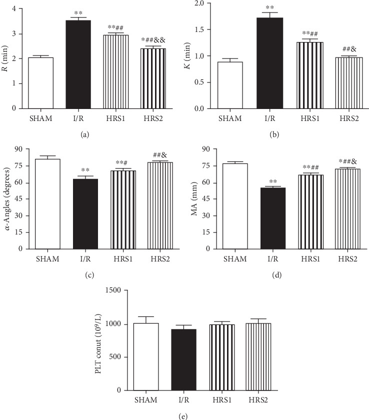
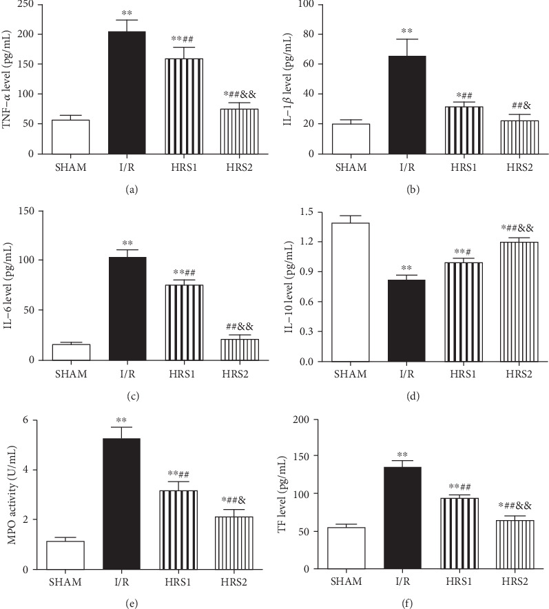
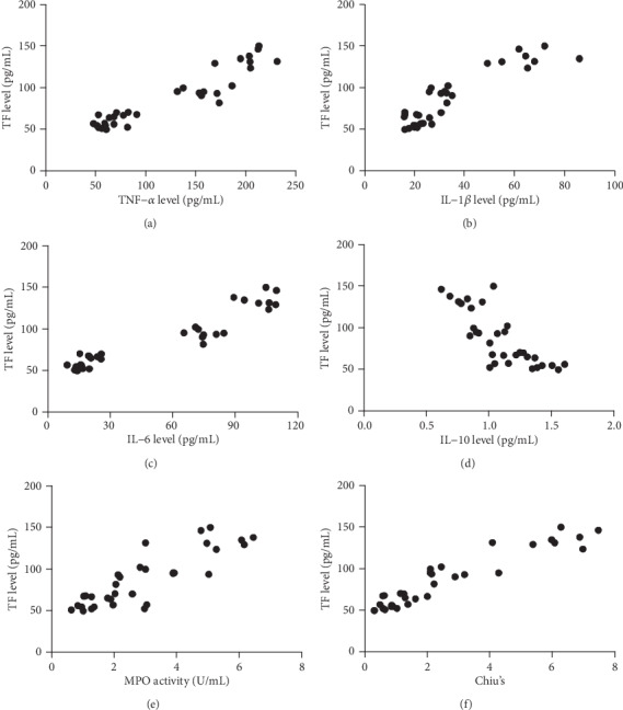
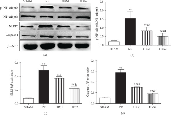
Intestinal ischemia/reperfusion (I/R) injury often causes inflammatory responses and coagulation disorders, which is further promoting the deterioration of the disease. Hydrogen has anti-inflammatory, antioxidative, and antiapoptotic properties against various diseases. However, the effect of hydrogen on coagulation dysfunction after intestinal I/R and the underlying mechanism remains unclear. The purpose of this study was to explore whether hydrogen-rich solution (HRS) could attenuate coagulation disorders and inflammation to improve intestinal injury and poor survival following intestinal I/R. The rat model of intestinal I/R injury was established by clamping the superior mesenteric artery for 90 min and reperfusion for 2 h. HRS (10 or 20 mL/kg) or 20 mL/kg 0.9% normal saline was intravenously injected at 10 min before reperfusion, respectively. The samples were harvested at 2 h after reperfusion for further analyses. Moreover, the survival rate was observed for 24 h. The results showed that HRS improved the survival rate and alleviated serum diamine oxidase activities, intestinal injury, edema, and apoptosis. Interestingly, HRS markedly improved intestinal I/R-mediated coagulation disorders as evidenced by abnormal conventional indicators of coagulation and thromboelastography. Additionally, HRS attenuated inflammatory responses and the elevated tissue factor (TF) and inhibited nuclear factor kappa beta (NF-κB) and nucleotide binding and oligomerization domain-like receptor family pyrin domain-containing 3 (NLRP3) inflammasome activation in peripheral blood mononuclear cells. Moreover, inflammatory factors and myeloperoxidase were closely associated with TF level. This study thus emphasized upon the amelioration of coagulation disorders and inflammation by HRS as a mechanism to improve intestinal I/R-induced intestinal injury and poor survival, which might be partially related to inhibition of NF-κB/NLRP3 pathway.


Conflict of interest statement

The authors declare that they have no conflicts of interest.

Figures

Figure 1
Figure 1
HRS improved the survival of rats after intestinal I/R. Rats subjected to intestinal ischemia for 90 minutes by occluding the superior mesenteric artery (SMA) were received normal saline (vehicle) or HRS (10 or 20 mL/kg of body weight) via the tail vein at 10 min before reperfusion and were observed for 24 hours. Data are presented as the survival percentage of animals. The survival rate is estimated and compared by the Kaplan-Meier method and the log-rank test (n = 15/group). P < 0.05, ∗∗P < 0.01 vs. SHAM group; #P < 0.05, ##P < 0.01 vs. I/R group; &P < 0.05 vs. HRS1 group.
Figure 2
Figure 2
HRS alleviated the histopathological damage in the intestine. (a) Representative images were shown for the pathological changes of intestinal mucosal tissues (hematoxylin and eosin staining, original magnification ×200). (b) The histopathological score of intestine (Chiu's score). (c) The W/D weight ratio. (d) Serum DAO activities. The data were represented as mean ± SD (n = 8/group). P < 0.05, ∗∗P < 0.01 vs. SHAM group; #P < 0.05, ##P < 0.01 vs. I/R group; &P < 0.05, &&P < 0.01 vs. HRS1 group. Scale bar = 50 μm.
Figure 3
Figure 3
HRS attenuated apoptosis in intestinal tissues. (a) Representative images for intestinal apoptotic cells (original magnification ×400). TUNEL-positive cells with dark brown nuclei indicated apoptosis (red arrows). (b) The quantitative analysis of apoptotic index among different groups. The data were represented as mean ± SD (n = 8/group). ∗∗P < 0.01 vs. SHAM group; ##P < 0.01 vs. I/R group; &&P < 0.01 vs. HRS1 group. Scale bar = 25 μm.
Figure 4
Figure 4
HRS improved coagulation disorders. (a) Prothrombin time (PT), (b) activated partial thrombin time (APTT), (c) thrombin time (TT), (d) fibrinogen (FIB), (e) fibrinogen degradation product (FDP), (f) d-dimer (D-Di), (g) prothrombin time-international standardization ratio (PT-INR). The data were represented as mean ± SD (n = 8/group). P < 0.05, ∗∗P < 0.01 vs. SHAM group; #P < 0.05, ##P < 0.01 vs. I/R group; &P < 0.05, &&P < 0.01 vs. HRS1 group.
Figure 5
Figure 5
Effects of HRS on thromboelastography (TEG) and platelet count. (a–e) were R, K, α-Angles, MA, and PLT count, respectively. The data were represented as mean ± SD (n = 8/group). P < 0.05, ∗∗P < 0.01 vs. SHAM group; #P < 0.05, ##P < 0.01 vs. I/R group; &P < 0.05, &&P < 0.01 vs. HRS1 group.
Figure 6
Figure 6
HRS reduced inflammatory stress and TF level. (a–f) were TNF-α, IL-1β, IL-6, IL-10, MPO, and TF levels, respectively. The data were represented as mean ± SD (n = 8/group). P < 0.05, ∗∗P < 0.01 vs. SHAM group; #P < 0.05, ##P < 0.01 vs. I/R group; &P < 0.05, &&P < 0.01 vs. HRS1 group.
Figure 7
Figure 7
Correlation analysis. Correlations (N = 32) between TF level and TNF-α level (a), TF level and IL-1β level (b), TF level and IL-6 level (c), TF level and IL-10 level (d), TF level and MPO activity (e), TF level and Chiu's score (f).
Figure 8
Figure 8
HRS inhibited NF-κB and NLRP3 inflammasome activation. Western blot was used to detect the expression levels of phospho-NF-κB p65, NF-κB p65, NLRP3, and Caspase 1 proteins in PBMCs (a–d). The data were represented as mean ± SD (n = 8/group). P < 0.05, ∗∗P < 0.01 vs. SHAM group; #P < 0.05, ##P < 0.01 vs. I/R group; &P < 0.05 vs. HRS1 group.

References

    1. Mallick I. H., Yang W., Winslet M. C., Seifalian A. M. Ischemia-reperfusion injury of the intestine and protective strategies against injury. Digestive diseases and sciences. 2004;49(9):1359–1377. doi: 10.1023/B:DDAS.0000042232.98927.91. - DOI - PubMed
    1. Grootjans J., Lenaerts K., Buurman W. A., Dejong C. H. C., Derikx J. P. M. Life and death at the mucosal-luminal interface: new perspectives on human intestinal ischemia-reperfusion. World journal of gastroenterology. 2016;22(9):2760–2770. doi: 10.3748/wjg.v22.i9.2760. - DOI - PMC - PubMed
    1. Zheng D. Y., Zhou M., Jin J., et al. Inhibition of P38 MAPK downregulates the expression of IL-1β to protect lung from acute injury in intestinal ischemia reperfusion rats. Mediators of inflammation. 2016;2016:8. doi: 10.1155/2016/9348037.9348037 - DOI - PMC - PubMed
    1. Fan X., Du J., Wang M.-H., et al. Irisin contributes to the hepatoprotection of dexmedetomidine during intestinal ischemia/reperfusion. Oxidative medicine and cellular longevity. 2019;2019:15. doi: 10.1155/2019/7857082.7857082 - DOI - PMC - PubMed
    1. Meng Q. T., Cao C., Wu Y., et al. Ischemic post-conditioning attenuates acute lung injury induced by intestinal ischemia-reperfusion in mice: role of Nrf2. Laboratory investigation. 2016;96(10):1087–1104. doi: 10.1038/labinvest.2016.87. - DOI - PubMed

MeSH terms

Substances