Case Reports
. 2020 Jun 10:11:451.
doi: 10.3389/fneur.2020.00451. eCollection 2020.

Classical Triad and Periventricular Lesions Do Not Necessarily Indicate Wernicke's Encephalopathy: A Case Report and Review of the Literature

Affiliations
Case Reports

Classical Triad and Periventricular Lesions Do Not Necessarily Indicate Wernicke's Encephalopathy: A Case Report and Review of the Literature

Lisha Ye et al. Front Neurol. .

Abstract

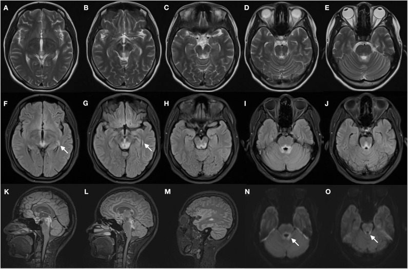
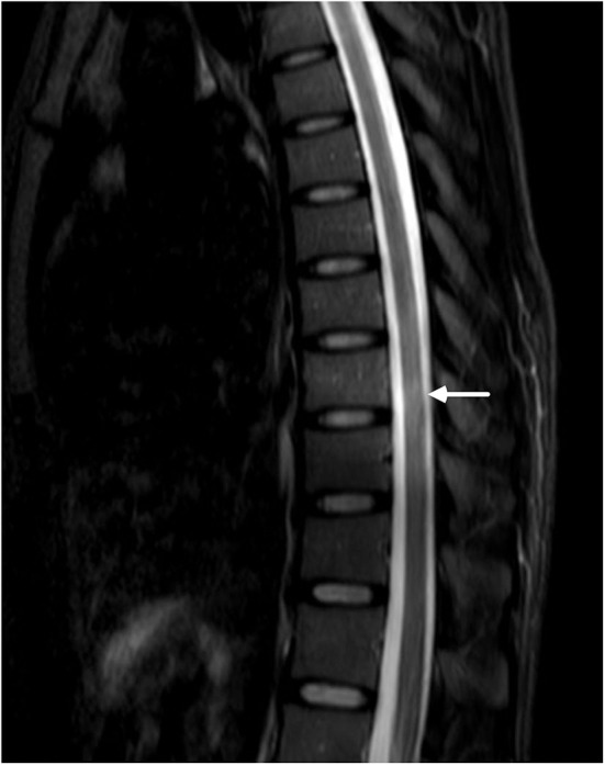
The classical triad-ophthalmoplegia, cerebellar dysfunction, and altered mental state-in addition to bilateral symmetrical periventricular lesions are actually common to see, and clinicians tend to associate that with Wernicke's encephalopathy (WE). The diagnosis is strengthened with a likely deficiency of thiamine. We herein describe a malnourished patient with clinical triad and hyperintensities in the circumventricular regions, and she turned out to have neuromyelitis optica spectrum disorder (NMOSD) after many twists and turns. Despite totally different pathogenic mechanisms, NMOSD can mimic WE, sometimes even exhibiting radiological features similar to that of WE, thereby complicating the diagnosis. Our case highlights how similar these two diseases could be and the importance of differential diagnosis in clinical practice, which are so far rarely reported. Some clinical and radiological differences of these two diseases are summarized to help establish a prompt diagnosis.

Keywords: Wernicke's encephalopathy; differential diagnosis; magnetic resonance imaging; neuromyelitis optica; periventricular lesions; triad.


Figures

Figure 1
Figure 1
Brain magnetic resonance images of the patient. T2-weighted images (A–E) demonstrate bilateral symmetrical hyperintense lesions in periaqueductal regions and around the fourth ventricle, which include superior colliculus (A), ventral tegmental area (B), inferior colliculus (C), and superior cerebellar peduncle (D,E). The abnormal regions revealed more obvious hyperintensity signal on corresponding FLAIR images (F–J), increased signal in the lateral ventricles was found (F,G, arrows). The FLAIR sagittal slice showed apparent hyperintense signal in the periaqueductal gray region (K) and surrounding the fourth ventricle (L), and lesions in the lateral ventricles (M). DWI images also manifest restricted water diffusion adjacent to the fourth ventricle (N,O, arrows).
Figure 2
Figure 2
MRI of the lower thoracic spine showed T2 hyperintense lesions from T7 through T9 (arrow). However, the signal was blurry, and we considered it to be undetermined.

Similar articles

Cited by

References

    1. Wingerchuk DM, Lennon VA, Lucchinetti CF, Pittock SJ, Weinshenker BG. The spectrum of neuromyelitis optica. Lancet Neurol. (2007) 6:805–15. 10.1016/S1474-4422(07)70216-8 - DOI - PubMed
    1. Reuler JB, Girard DE, Cooney TG. Current concepts. Wernicke's encephalopathy. N Engl J Med. (1985) 312:1035–9. 10.1056/NEJM198504183121606 - DOI - PubMed
    1. Shan F, Zhong R, Wu L, Fan Y, Long Y, Gao C. Neuromyelitis optica spectrum disorders may be misdiagnosed as Wernicke's encephalopathy. Int J Neurosci. (2016) 126:922–7. 10.3109/00207454.2015.1084619 - DOI - PubMed
    1. Rash JE, Yasumura T, Hudson CS, Agre P, Nielsen S. Direct immunogold labeling of aquaporin-4 in square arrays ofastrocyte and ependymocyte plasma membranes in rat brain and spinalcord. Proc Natl Acad Sci USA. (1998) 95:11981–6. 10.1073/pnas.95.20.11981 - DOI - PMC - PubMed
    1. Papadopoulos MC, Verkman A. Aquaporin 4 and neuromyelitis optica. Lancet Neurol. (2012) 11:535–44. 10.1016/S1474-4422(12)70133-3 - DOI - PMC - PubMed

Publication types

LinkOut - more resources