A Single Amino Acid Substitution in the Matrix Protein (M51R) of Vesicular Stomatitis New Jersey Virus Impairs Replication in Cultured Porcine Macrophages and Results in Significant Attenuation in Pigs
- PMID: 32587580
- PMCID: PMC7299242
- DOI: 10.3389/fmicb.2020.01123
A Single Amino Acid Substitution in the Matrix Protein (M51R) of Vesicular Stomatitis New Jersey Virus Impairs Replication in Cultured Porcine Macrophages and Results in Significant Attenuation in Pigs
Abstract
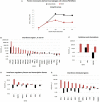
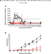
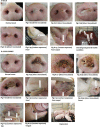
In this study, we explore the virulence of vesicular stomatitis New Jersey virus (VSNJV) in pigs and its potential relationship with the virus's ability to modulate innate responses. For this purpose, we developed a mutant of the highly virulent strain NJ0612NME6, containing a single amino acid substitution in the matrix protein (M51R). The M51R mutant of NJ0612NME6 was unable to suppress the transcription of genes associated with the innate immune response both in primary fetal porcine kidney cells and porcine primary macrophage cultures. Impaired viral growth was observed only in porcine macrophage cultures, indicating that the M51 residue is required for efficient replication of VSNJV in these cells. Furthermore, when inoculated in pigs by intradermal scarification of the snout, M51R infection was characterized by decreased clinical signs including reduced fever and development of less and smaller secondary vesicular lesions. Pigs infected with M51R had decreased levels of viral shedding and absence of RNAemia compared to the parental virus. The ability of the mutant virus to infect pigs by direct contact remained intact, indicating that the M51R mutation resulted in a partially attenuated phenotype capable of causing primary lesions and transmitting to sentinel pigs. Collectively, our results show a positive correlation between the ability of VSNJV to counteract the innate immune response in swine macrophage cultures and the level of virulence in pigs, a natural host of this virus. More studies are encouraged to evaluate the interaction of VSNJV with macrophages and other components of the immune response in pigs.
Keywords: M51R; immune response; macrophages; pathogenesis; type 1 interferon; vesicular stomatitis; virulence.
Copyright © 2020 Velazquez-Salinas, Pauszek, Holinka, Gladue, Rekant, Bishop, Stenfeldt, Verdugo-Rodriguez, Borca, Arzt and Rodriguez.
Figures


Similar articles
-
Exploring the Molecular Basis of Vesicular Stomatitis Virus Pathogenesis in Swine: Insights from Expression Profiling of Primary Macrophages Infected with M51R Mutant Virus.Pathogens. 2023 Jun 30;12(7):896. doi: 10.3390/pathogens12070896. Pathogens. 2023. PMID: 37513744 Free PMC article.
-
Infection of guinea pigs with vesicular stomatitis New Jersey virus Transmitted by Culicoides sonorensis (Diptera: Ceratopogonidae).J Med Entomol. 2006 May;43(3):568-73. doi: 10.1603/0022-2585(2006)43[568:iogpwv]2.0.co;2. J Med Entomol. 2006. PMID: 16739417
-
Increased Virulence of an Epidemic Strain of Vesicular Stomatitis Virus Is Associated With Interference of the Innate Response in Pigs.Front Microbiol. 2018 Aug 15;9:1891. doi: 10.3389/fmicb.2018.01891. eCollection 2018. Front Microbiol. 2018. PMID: 30158915 Free PMC article.
-
Creation of matrix protein gene variants of two serotypes of vesicular stomatitis virus as prime-boost vaccine vectors.J Virol. 2015 Jun;89(12):6338-51. doi: 10.1128/JVI.00222-15. Epub 2015 Apr 8. J Virol. 2015. PMID: 25855732 Free PMC article.
-
Matrix protein of VSV New Jersey serotype containing methionine to arginine substitutions at positions 48 and 51 allows near-normal host cell gene expression.Virology. 2007 Jan 5;357(1):41-53. doi: 10.1016/j.virol.2006.07.022. Epub 2006 Sep 7. Virology. 2007. PMID: 16962155
Cited by
-
Phenotypic Differences Between the Epidemic Strains of Vesicular Stomatitis Virus Serotype Indiana 98COE and IN0919WYB2 Using an In-Vivo Pig (Sus scrofa) Model.Viruses. 2024 Dec 13;16(12):1915. doi: 10.3390/v16121915. Viruses. 2024. PMID: 39772222 Free PMC article.
-
Comparison of Endemic and Epidemic Vesicular Stomatitis Virus Lineages in Culicoides sonorensis Midges.Viruses. 2022 Jun 3;14(6):1221. doi: 10.3390/v14061221. Viruses. 2022. PMID: 35746691 Free PMC article.
-
Molecular Pathogenesis and Immune Evasion of Vesicular Stomatitis New Jersey Virus Inferred from Genes Expression Changes in Infected Porcine Macrophages.Pathogens. 2021 Sep 3;10(9):1134. doi: 10.3390/pathogens10091134. Pathogens. 2021. PMID: 34578166 Free PMC article.
-
Exploring the Molecular Basis of Vesicular Stomatitis Virus Pathogenesis in Swine: Insights from Expression Profiling of Primary Macrophages Infected with M51R Mutant Virus.Pathogens. 2023 Jun 30;12(7):896. doi: 10.3390/pathogens12070896. Pathogens. 2023. PMID: 37513744 Free PMC article.
-
Molecular Tracking of the Origin of Vesicular Stomatitis Outbreaks in 2004 and 2018, Ecuador.Vet Sci. 2023 Feb 24;10(3):181. doi: 10.3390/vetsci10030181. Vet Sci. 2023. PMID: 36977220 Free PMC article.
References
-
- Ahmed M., Mckenzie M. O., Puckett S., Hojnacki M., Poliquin L., Lyles D. S. (2003). Ability of the matrix protein of vesicular stomatitis virus to suppress beta interferon gene expression is genetically correlated with the inhibition of host RNA and protein synthesis. J. Virol. 77, 4646–4657. 10.1128/JVI.77.8.4646-4657.2003, PMID: - DOI - PMC - PubMed
-
- Arzt J., Pacheco J. M., Rodriguez L. L. (2010). The early pathogenesis of foot-and-mouth disease in cattle after aerosol inoculation. Identification of the nasopharynx as the primary site of infection. Vet Pathol. 47, 1048–1063. - PubMed
LinkOut - more resources
Full Text Sources

