

Reliability of Endoscopic Ultrasound Using Miniprobes and Grayscale Histogram Analysis in Diagnosing Upper Gastrointestinal Subepithelial Lesions
- PMID: 32587613
- PMCID: PMC7301246
- DOI: 10.1155/2020/6591341
Reliability of Endoscopic Ultrasound Using Miniprobes and Grayscale Histogram Analysis in Diagnosing Upper Gastrointestinal Subepithelial Lesions
Abstract
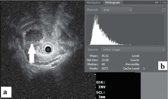
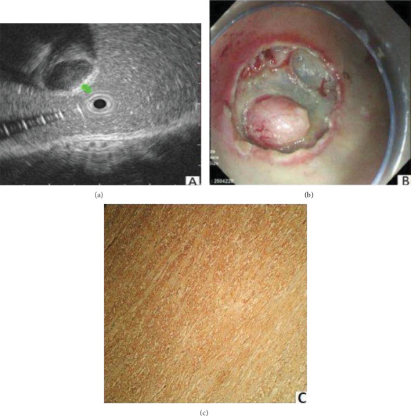
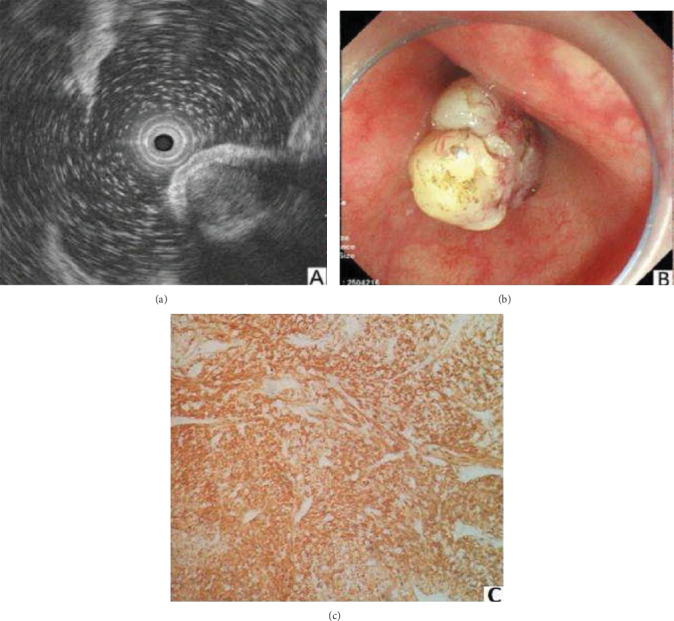
Background: To assess the role of endoscopic ultrasound (EUS) in the diagnosis of upper gastrointestinal subepithelial lesions (SELs) and to investigate EUS combined with a grayscale histogram analysis for the differentiation of leiomyomas and gastrointestinal stromal tumors (GISTs).
Methods: A retrospective study of 709 patients with upper gastrointestinal SELs was conducted by EUS before endoscopic resection. The EUS findings of SELs and pathological results after endoscopic resection were compared. The EUS images of SELs, particularly, leiomyoma and GIST, were further analyzed via a grayscale histogram to differentiate between the two tumors.
Results: Of the 709 patients, 47 cases were pathologically undetermined. The diagnostic consistency of EUS with endoscopic resection was 88.2% (584/662), including 185 muscularis mucosa, 61 submucosa, and 338 muscularis propria, respectively. The diagnostic consistency of EUS with pathology was 80.1% (530/662). The gray value of GISTs was significantly higher than that of leiomyomas (58.9 ± 8.3 vs. 39.5 ± 5.9, t = 57.0, P < 0.0001). The standard deviation of leiomyomas was significantly lower than that of GISTs (20.6 ± 7.0 vs. 39.8 ± 9.3, t = 23.7, P < 0.0001). The grayscale histogram analysis of GISTs showed higher echo ultrasound, and the echo of leiomyoma was more uniform.
Conclusion: EUS is the preferred procedure for the evaluation of upper gastrointestinal SELs. EUS combined with a grayscale histogram analysis is an effective method for the differentiation of leiomyomas and GISTs.
Copyright © 2020 Samiullah Khan et al.
Conflict of interest statement
The authors have no conflicts of interest to declare.
Figures
References
LinkOut - more resources
Full Text Sources
