. 2020 Jun 10:2020:4242130.
doi: 10.1155/2020/4242130. eCollection 2020.

Human Adipose Derived Cells in Two- and Three-Dimensional Cultures: Functional Validation of an In Vitro Fat Construct

Affiliations

Human Adipose Derived Cells in Two- and Three-Dimensional Cultures: Functional Validation of an In Vitro Fat Construct

Robert Bender et al. Stem Cells Int. .

Abstract

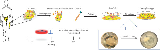
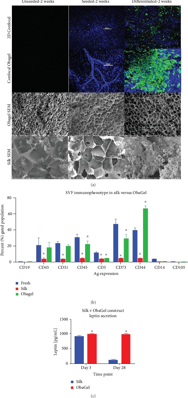
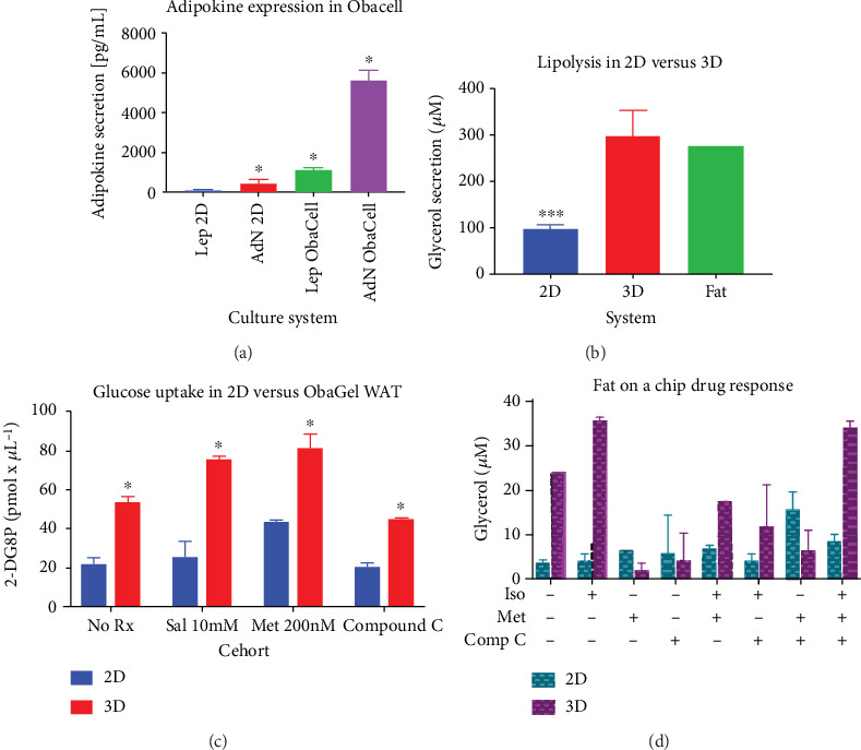
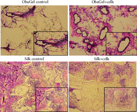
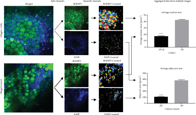
Obesity, defined as a body mass index of 30 kg/m2 or above, has increased considerably in incidence and frequency within the United States and globally. Associated comorbidities including cardiovascular disease, type 2 diabetes mellitus, metabolic syndrome, and nonalcoholic fatty liver disease have led to a focus on the mechanisms promoting the prevention and treatment of obesity. Commonly utilized in vitro models employ human or mouse preadipocyte cell lines in a 2-dimensional (2D) format. Due to the structural, biochemical, and biological limitations of these models, increased attention has been placed on "organ on a chip" technologies for a 3-dimensional (3D) culture. Herein, we describe a method employing cryopreserved primary human stromal vascular fraction (SVF) cells and a human blood product-derived biological scaffold to create a 3D adipose depot in vitro. The "fat-on-chip" 3D cultures have been validated relative to 2D cultures based on proliferation, flow cytometry, adipogenic differentiation, confocal microscopy/immunofluorescence, and functional assays (adipokine secretion, glucose uptake, and lipolysis). Thus, the in vitro culture system demonstrates the critical characteristics required for a humanized 3D white adipose tissue (WAT) model.


Conflict of interest statement

The following authors state the following conflicts of interest: A. Alarcon, T. Frazier, J. M. Gimble, and X. Wu are all employees of LaCell LLC and Obatala Sciences Inc.; T. Frazier, J.M. Gimble, and X. Wu are cofounders and coowners of Obatala Sciences; J.M. Gimble and X. Wu are coowners and cofounders of LaCell. All remaining authors have no conflicts to declare.

Figures

Figure 1
Figure 1
Summary figure representing the overall process for developing static fat-on-a-chip cultures using ObaGel.
Figure 2
Figure 2
Demographic characteristics of donors and SVF cells utilized in the study. Human SVF cells were characterized on the basis of (a) adipogenic and osteogenic differentiation, (b) donor demographics, (c) colony formation, (d) proliferation, and (e) cell immunophenotype. Donor demographics are as follows (in mean ± SD): age (49.4 ± 6.3), body mass index (BMI 27.5 ± 2.9).
Figure 3
Figure 3
Temporal immunophenotype and gelling behavior of SVF cells cultured in 3 dimensions in human-derived ObaGel. Studies investigating the correlation of cell seeding density and ObaGel concentration to gel stability were performed (a), and whole plate images were taken (b). Gel stability was measured and reported as a percentage (%) total gelling. Investigations into impact on SVF immunophenotype from freshly isolated SVF cells compared to ObaGel cultures at days 1 and 7 (c). 100% gel formation was maintained in 7.5% ObaGel-supplemented medium (v/v), in the absence of any additional supplementary growth factors (d). Values reported as mean ± SD. p < 0.05.
Figure 4
Figure 4
Comparison of SVF proliferation, spheroid volume and formation, and adipogenic functionality in 2D culture and ObaGel. (a) Bright field and confocal microscopy images of SVF cell proliferation in 2D culture and ObaGel. Cells were seeded in traditional 2D media or ObaGel, cultured for 5 days, and stained with the Hoechst dye. Robust cell proliferation occurs in ObaGel compared to 2D culture, as indicated by Hoechst positivity. (b, c) report photomicrographs and respective quantification that reflect a positive correlation between ObaGel concentration and SVF spheroid size, density, and volume within 5-7 days of culture. (d) demonstrates that adipogenesis occurs within spheroids as early as day 3 of differentiation. Values reported as mean ± SD. p < 0.05 when compared to the freshly thawed cells or silk constructs.
Figure 5
Figure 5
A comparison of the in vitro behavior of human SVF cells when cultured in 2-dimensional conditions and in 3 dimensions when seeded on either silk scaffolds or cultured in ObaGel for 2 weeks. (a) Confocal and cryoscanning electron microscopy (cryo-SEM) images of SVF cell seeding, proliferation, and adipogenic differentiation behavior in traditional 2D culture, silk scaffolds, and ObaGel. (b) SVF immunophenotype following 1 week of culture reveals that ObaGel is superior in maintaining a heterogenic immunophenotype similar to freshly isolated cells and supports the expansion of CD44-positive subpopulations (b), and adipokine functionality (c) reports the measure of leptin secretion from adipogenic differentiated cells seeded on silk or with ObaGel are early (day 3) and late (day 28) time points of differentiation. Immunophenotype values reported as mean (μ) ± standard deviation (SD). p < 0.05.
Figure 6
Figure 6
ObaGel cultures exhibit physiologically relevant functionality. Adiponectin (AdN) and leptin (Lep) secretions were measured from traditional cell cultures (2D) compared to three-dimensional adipose cultures (ObaCell) after 21 days of culture. Values reported are in pg/mL conditioned medium and are reported as mean (μ) ± standard deviation (SD). p < 0.05. Adipose tissue functionality demonstrated with measurement of glycerol secretion in a conditioned medium from cells differentiated in two dimensions (2D) compared to ObaGel after 14 days of culture. ObaGel cultures demonstrated higher sensitivity to AMPK-targeting compounds isoproterenol, metformin, compound C, and IBMX. Values reported are in pg/mL conditioned medium and are reported as mean (μ) ± standard deviation (SD). p < 0.05.
Figure 7
Figure 7
Implantation of ObaGel for the development of a humanized fat pad in mice. Scaffolds remained in mice for 6 weeks. Hematoxylin and eosin staining revealed a dense confinement of proliferating cells within the silk scaffolds. In contrast, ObaGel implants contained large blood vessels that were clearly integrated functionally within larger, more mature adipocytes.
Figure 8
Figure 8
In vivo analysis of implants of human SVF cell ObaGel constructs in immunodeficient mice. BODIPY lipophilic staining was performed in explants removed from nude mice following 6-week implantation. Images were taken by confocal laser microscopy and quantified using CellProfiler v2.2.0 software. Values reported are in pg/mL conditioned medium and are reported as mean (μ) ± standard deviation (SD). p < 0.05.

Similar articles

Cited by

References

    1. Bray G. A., Kim K. K., Wilding J. P. H., on behalf of the World Obesity Federation Obesity: a chronic relapsing progressive disease process. A position statement of the World Obesity Federation. Obesity Reviews. 2017;18(7):715–723. doi: 10.1111/obr.12551. - DOI - PubMed
    1. Hauner H., Entenmann G., Wabitsch M., et al. Promoting effect of glucocorticoids on the differentiation of human adipocyte precursor cells cultured in a chemically defined medium. The Journal of Clinical Investigation. 1989;84(5):1663–1670. doi: 10.1172/JCI114345. - DOI - PMC - PubMed
    1. Green H., Kehinde O. An established preadipose cell line and its differentiation in culture II. Factors affecting the adipose conversion. Cell. 1975;5(1):19–27. doi: 10.1016/0092-8674(75)90087-2. - DOI - PubMed
    1. Bourin P., Bunnell B. A., Casteilla L., et al. Stromal cells from the adipose tissue-derived stromal vascular fraction and culture expanded adipose tissue-derived stromal/stem cells: a joint statement of the International Federation for Adipose Therapeutics and Science (IFATS) and the International Society for Cellular Therapy (ISCT) Cytotherapy. 2013;15(6):641–648. doi: 10.1016/j.jcyt.2013.02.006. - DOI - PMC - PubMed
    1. Sadie-Van Gijsen H. Adipocyte biology: it is time to upgrade to a new model. Journal of Cellular Physiology. 2019;234(3):2399–2425. doi: 10.1002/jcp.27266. - DOI - PubMed

LinkOut - more resources