. 2020 May 15;17(2):328-342.
doi: 10.20892/j.issn.2095-3941.2019.0229.

CDK11 negatively regulates Wnt/β-catenin signaling in the endosomal compartment by affecting microtubule stability

Affiliations

CDK11 negatively regulates Wnt/β-catenin signaling in the endosomal compartment by affecting microtubule stability

Danmin Ou et al. Cancer Biol Med. .

Abstract

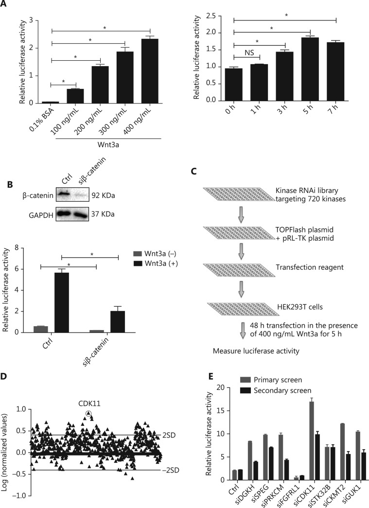
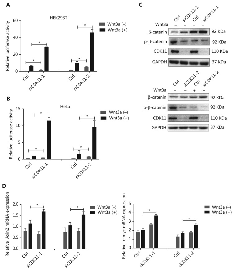
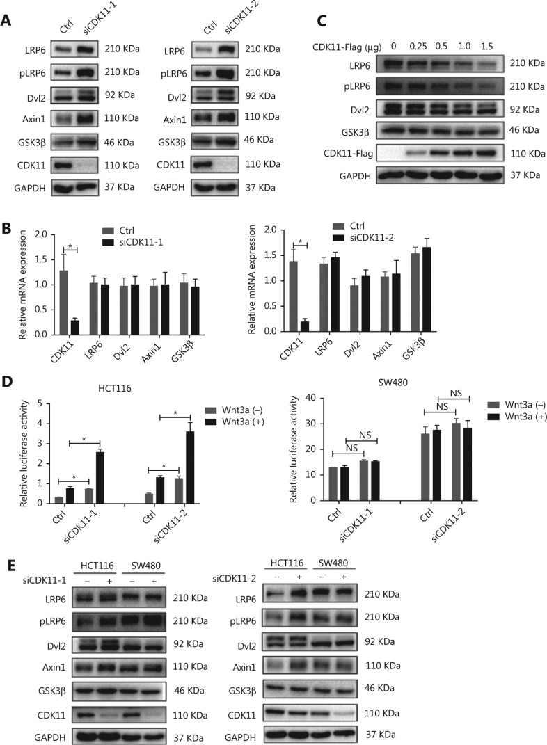
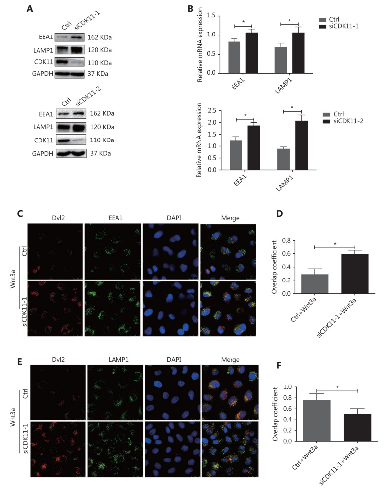
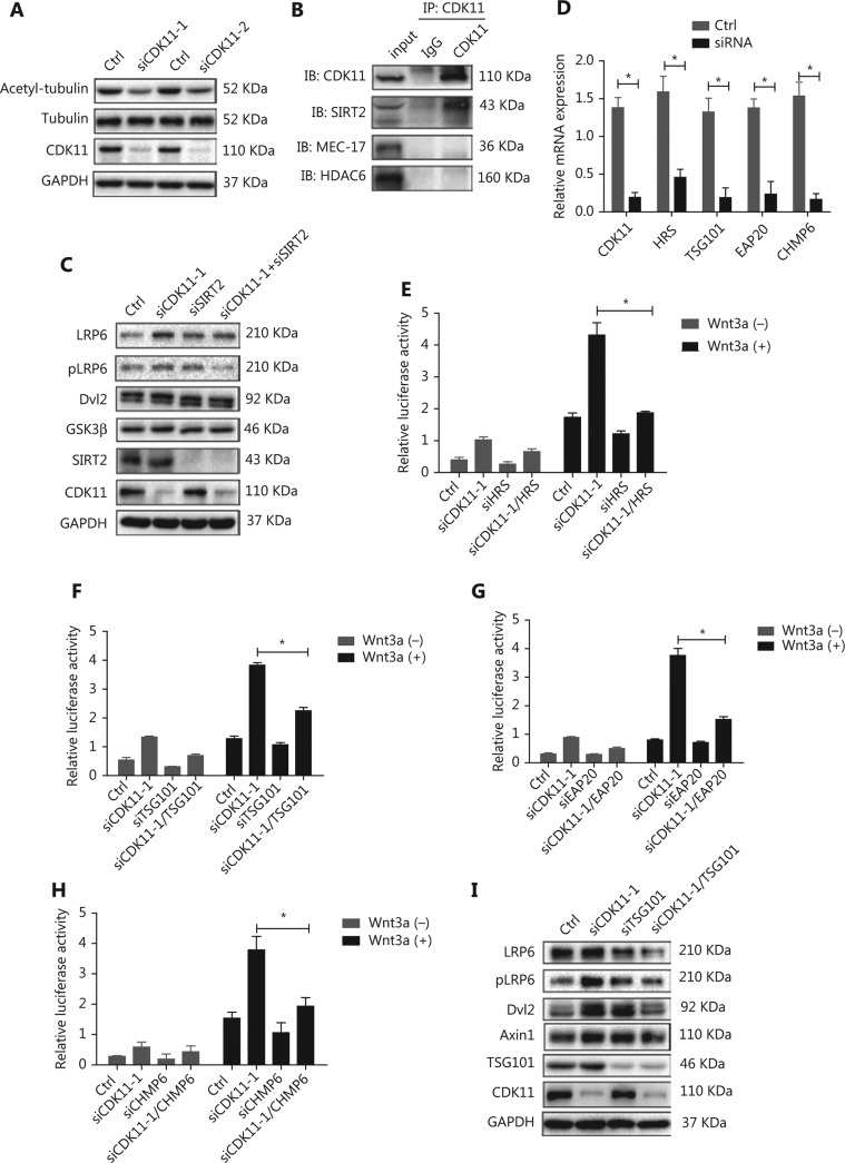
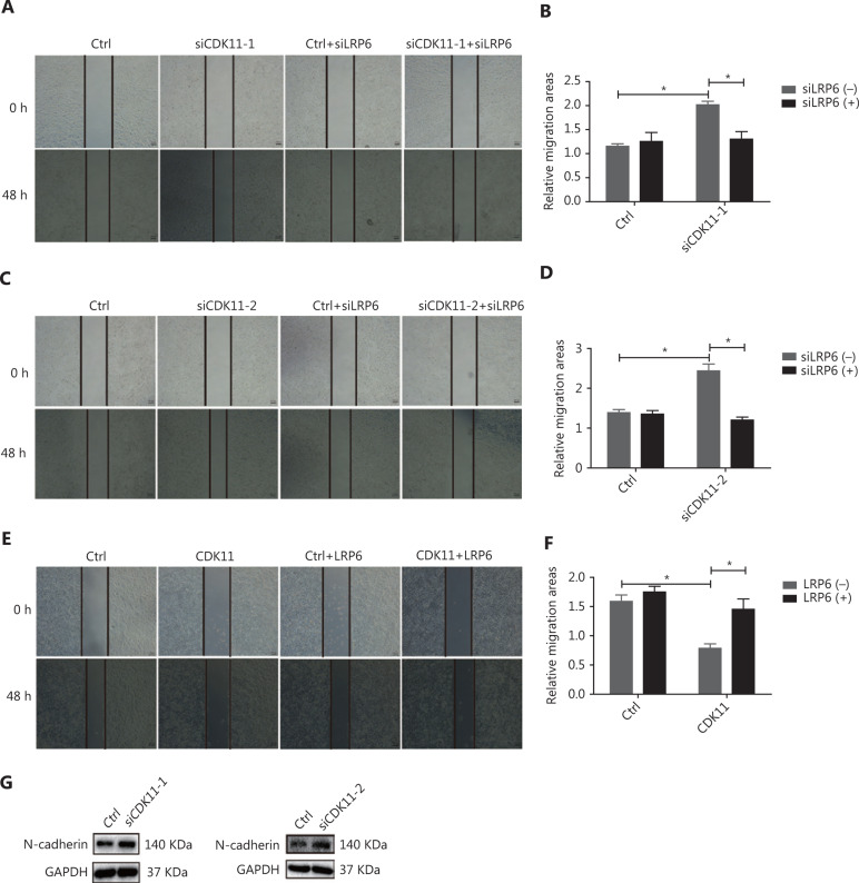
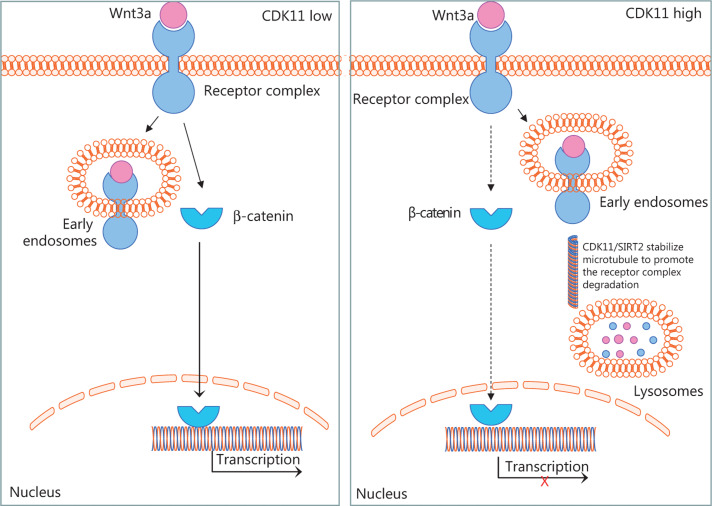
Objectives: Improper activation of Wnt/β-catenin signaling has been implicated in human diseases. Beyond the well-studied glycogen synthase kinase 3β (GSK3β) and casein kinase 1 (CK1), other kinases affecting Wnt/β-catenin signaling remain to be defined. Methods:To identify the kinases that modulate Wnt/β-catenin signaling, we applied a kinase small interfering RNA (siRNA) library screen approach. Luciferase assays, immunoblotting, and real-time polymerase chain reaction (PCR) were performed to confirm the regulation of the Wnt/β-catenin signaling pathway by cyclin-dependent kinase 11 (CDK11) and to investigate the underlying mechanism. Confocal immunofluorescence, coimmunoprecipitation (co-IP), and scratch wound assays were used to demonstrate colocalization, detect protein interactions, and explore the function of CDK11. Results: CDK11 was found to be a significant candidate kinase participating in the negative control of Wnt/β-catenin signaling. Down-regulation of CDK11 led to the accumulation of Wnt/β-catenin signaling receptor complexes, in a manner dependent on intact adenomatosis polyposis coli (APC) protein. Further analysis showed that CDK11 modulation of Wnt/β-catenin signaling engaged the endolysosomal machinery, and CDK11 knockdown enhanced the colocalization of Wnt/β-catenin signaling receptor complexes with early endosomes and decreased colocalization with lysosomes. Mechanistically, CDK11 was found to function in Wnt/β-catenin signaling by regulating microtubule stability. Depletion of CDK11 down-regulated acetyl-α-tubulin. Moreover, co-IP assays demonstrated that CDK11 interacts with the α-tubulin deacetylase SIRT2, whereas SIRT2 down-regulation in CDK11-depleted cells reversed the accumulation of Wnt/β-catenin signaling receptor complexes. CDK11 was found to suppress cell migration through altered Wnt/β-catenin signaling. Conclusions: CDK11 is a negative modulator of Wnt/β-catenin signaling that stabilizes microtubules, thus resulting in the dysregulation of receptor complex trafficking from early endosomes to lysosomes.

Keywords: CDK11; SIRT2; Wnt/β-catenin signaling; endosome; microtubule.


Figures

Figure 1
Figure 1
RNAi screen of kinases regulating Wnt/β-catenin signaling activity. (A) Relative luciferase activity in HEK293T cells cotransfected with TOPFlash and pRL-TK plasmids under Wnt3a stimulation at different doses and time points (*P < 0.05; NS, no statistical significance). (B) Relative luciferase activity in HEK293T cells cotransfected with luciferase reporter plasmids and nontargeting siRNA or β-catenin siRNA with or without Wnt3a stimulation. Top, results of β-catenin depletion (*P < 0.05). (C) Schematic of the RNAi screen strategy. (D) Log ratio of the relative luciferase activity of targeting kinase siRNAs to nontargeting siRNAs, under Wnt3a stimulation, in HEK293T cells. Two SD of the log ratios of 720 candidates were quantified and are indicated in the figure. (E) Secondary screen for Wnt/β-catenin signaling activity of 8 representative kinases in HEK293T cells was performed and is shown with the primary screen results. All results are representative of 3 independent experiments.
Figure 2
Figure 2
Negative regulation of Wnt/β-catenin signaling by CDK11. (A) Relative luciferase activity in CDK11-depleted HEK293T cells with or without Wnt3a stimulation (*P < 0.05). (B) Relative luciferase activity in CDK11-depleted HeLa cells with or without Wnt3a stimulation (*P < 0.05). (C) Immunoblotting analysis of β-catenin and phospho-β-catenin (Ser33/Ser37/Thr41) expression in CDK11-depleted HeLa cells with or without Wnt3a stimulation. (D) mRNA levels of Axin2 and c-myc measured by real-time polymerase chain reaction (PCR) in CDK11-depleted HeLa cells with or without Wnt3a stimulation (*P < 0.05). All results are representative of 3 independent experiments.
Figure 3
Figure 3
Wnt/β-catenin signaling components accumulate after CDK11 depletion in an intact APC-dependent manner. (A) Western blot analysis of LRP6, pLRP6, Dvl2, Axin1, and GSK3β expression in CDK11-depleted HeLa cells. (B) mRNA levels of LRP6, Dvl2, Axin1, and GSK3β, measured by real-time PCR in CDK11-depleted HeLa cells (*P < 0.05). (C) Western blot analysis of LRP6, pLRP6, Dvl2, and GSK3β expression in CDK11-overexpressing HeLa cells. (D) Relative luciferase activity in CDK11-depleted HCT116 and SW480 cells with or without Wnt3a stimulation (*P < 0.05; NS, no statistical significance). (E) Western blot analysis of LRP6, pLRP6, Dvl2, Axin1, and GSK3β expression in CDK11-depleted HCT116 and SW480 cells. All results are representative of 3 independent experiments.
Figure 4
Figure 4
Receptor complexes are stuck in early endosomes in CDK11-depleted cells. (A) Western blot analysis was performed to examine the expression of EEA1 and LAMP1 in CDK11-depleted HeLa cells. (B) Real-time PCR was performed to examine the mRNA levels of EEA1 and LAMP1 in CDK11-depleted HeLa cells (*P < 0.05). (C) In control and CDK11-depleted HeLa cells stimulated with Wnt3a, confocal immunofluorescence assays showed the colocalization of Dvl2 with EEA1 (400×). (D) Quantification of the overlap coefficient of Dvl2 with EEA1 in control and CDK11-depleted cells stimulated with Wnt3a (*P < 0.05). (E) In control and CDK11-depleted HeLa cells stimulated with Wnt3a, confocal immunofluorescence assays showed the colocalization of Dvl2 with LAMP1 (400×). (F) Quantification of the overlap coefficient of Dvl2 with LAMP1 in control and CDK11-depleted cells stimulated with Wnt3a (*P < 0.05). All results are representative of 3 independent experiments.
Figure 5
Figure 5
Disruption of microtubule stability and ESCRT reverses the modulation of CDK11 on Wnt/β-catenin signaling. (A) Western blot analysis of acetyl-α-tubulin expression in CDK11-depleted HeLa cells. (B) Co-IP assays were performed to examine the interactions among CDK11 and SIRT2, HDAC6, and MEC-17 in HeLa cells. (C) Western blot analysis of the receptor complex levels of Wnt/β-catenin signaling in CDK11- and SIRT2-depleted HeLa cells. (D) Real-time PCR was performed to examine the expression of CDK11, HRS, TSG101, EAP20, and CHMP6 after knockdown of CDK11, HRS, TSG101, EAP20, and CHMP6, respectively, in HeLa cells (*P < 0.05). (E-H) Luciferase reporter assays showing the effect of ESCRT complex depletion on the activation of Wnt/β-catenin signaling induced by CDK11 depletion in HeLa cells (*P < 0.05). (I) Protein levels of LRP6, pLRP6, Dvl2, and Axin1 were examined in CDK11- and TSG101-depleted HeLa cells. All results are representative of 3 independent experiments.
Figure 6
Figure 6
CDK11 depletion promotes migration through Wnt/β-catenin signaling. (A-D) Representative images (50×; A and C) and quantification (B and D) of the scratch wound assay results in CDK11- and LRP6-depleted HeLa cells (*P < 0.05). (E and F) Representative images (50×; E) and quantification (F) of the scratch wound assay results in CDK11- and LRP6-overexpressing HeLa cells (*P < 0.05). (G) Western blot analysis of N-cadherin expression in CDK11-depleted HeLa cells. All results are representative of 3 independent experiments.
Figure 7
Figure 7
Schematic of CDK11 regulation of Wnt/β-catenin signaling, according to our results. The diagram shows that CDK11 regulates the trafficking of Wnt/β-catenin signaling receptor complexes between early endosomes and lysosomes by modulating microtubule stability. When CDK11 is present at a low level, the receptor complexes are retained in early endosomes, and Wnt/β-catenin signaling is active. When CDK11 is present at a high level, microtubule stability is enhanced, and the receptor complexes traffic from early endosomes to lysosomes for degradation increases, thus leading to the inactivation of Wnt/β-catenin signaling.

Similar articles

Cited by

References

    1. Clevers H. Wnt/beta-catenin signaling in development and disease. Cell. 2006;127:469–80. - PubMed
    1. Polakis P. The many ways of Wnt in cancer. Curr Opin Genet Dev. 2007;17:45–51. - PubMed
    1. Anastas JN, Moon RT. Wnt signalling pathways as therapeutic targets in cancer. Nat Rev Cancer. 2013;13:11–26. - PubMed
    1. Aberle H, Bauer A, Stappert J, Kispert A, Kemler R. Beta-catenin is a target for the ubiquitin-proteasome pathway. EMBO J. 1997;16:3797–804. - PMC - PubMed
    1. Li VS, Ng SS, Boersema PJ, Low TY, Karthaus WR, Gerlach JP, et al. Wnt signaling through inhibition of beta-catenin degradation in an intact Axin1 complex. Cell. 2012;149:1245–56. - PubMed

Publication types