. 2020 May 15;17(2):387-400.
doi: 10.20892/j.issn.2095-3941.2019.0380.

DYRK1A suppression restrains Mcl-1 expression and sensitizes NSCLC cells to Bcl-2 inhibitors

Affiliations

DYRK1A suppression restrains Mcl-1 expression and sensitizes NSCLC cells to Bcl-2 inhibitors

Yangling Li et al. Cancer Biol Med. .

Abstract

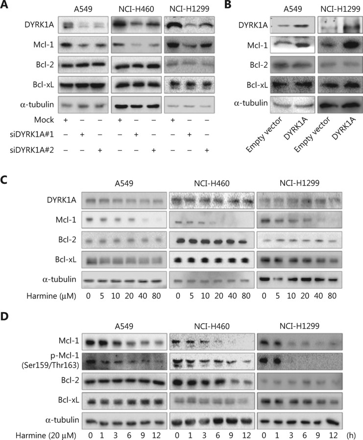
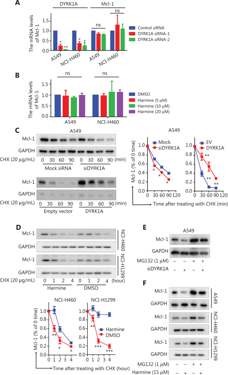
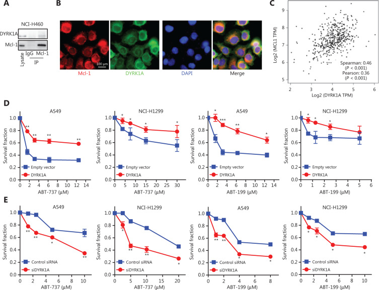
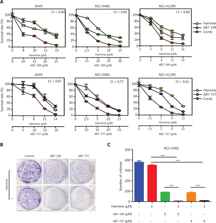
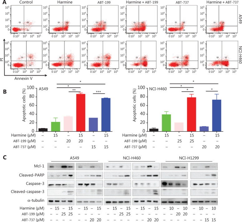
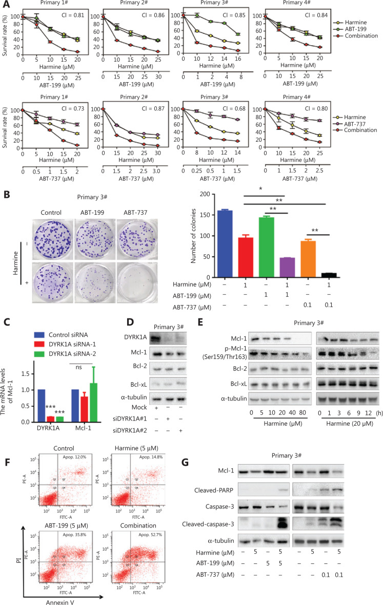
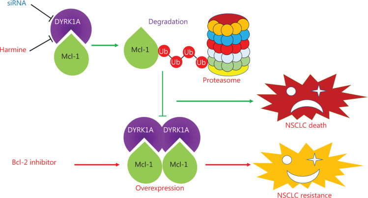
Objective: Mcl-1 overexpression confers acquired resistance to Bcl-2 inhibitors in non-small cell lung cancer (NSCLC), but no direct Mcl-1 inhibitor is currently available for clinical use. Thus, novel therapeutic strategies are urgently needed to target Mcl-1 and sensitize the anti-NSCLC activity of Bcl-2 inhibitors. Methods: Cell proliferation was measured using sulforhodamine B and colony formation assays, and apoptosis was detected with Annexin V-FITC staining. Gene expression was manipulated using siRNAs and plasmids. Real-time PCR and Western blot were used to measure mRNA and protein levels. Immunoprecipitation and immunofluorescence were used to analyze co-localization of dual specificity tyrosine-phosphorylation-regulated kinase 1A (DYRK1A) and Mcl-1. Results: Suppression of DYRK1A resulted in reduced Mcl-1 expression in NSCLC cells, whereas overexpression of DYRK1A significantly increased Mcl-1 expression. Suppression of DYRK1A did not alter Mcl-1 mRNA levels, but did result in an accelerated degradation of Mcl-1 protein in NSCLC cells. Furthermore, DYRK1A mediated proteasome-dependent degradation of Mcl-1 in NSCLC cells, and DYRK1A co-localized with Mcl-1 in NSCLC cells and was co-expressed with Mcl-1 in tumor samples from lung cancer patients, suggesting that Mcl-1 may be a novel DYRK1A substrate. We showed that combined therapy with harmine and Bcl-2 antagonists significantly inhibited cell proliferation and induced apoptosis in NSCLC cell lines as well as primary NSCLC cells. Conclusions: Mcl-1 is a novel DYRK1A substrate, and the role of DYRK1A in promoting Mcl-1 stability makes it an attractive target for decreasing Bcl-2 inhibitor resistance.

Keywords: Bcl-2 inhibitor; DYRK1A; Mcl-1; NSCLC; combination.


Figures

Figure 1
Figure 1
DYRK1A regulates the expression of Mcl-1 in NSCLC cells. (A) NSCLC cells were transfected with control siRNA and DYRK1A siRNA for 48 h, and the expression of DYRK1A and Bcl-2 family members were detected by Western blot. (B) NSCLC cells were transfected with empty vector or DYRK1A plasmid for 48 h, and the expression of DYRK1A and Bcl-2 family members were detected by Western blot. (C) NSCLC cells were treated with harmine at the indicated concentrations for 24 h, and the expression of the indicated proteins were detected by Western blot. (D) NSCLC cells were treated with 20 μM harmine for 1, 3, 6, 9, and 12 h, and the expression of the indicated proteins was detected by Western blot.
Figure 2
Figure 2
DYRK1A promotes the stability of Mcl-1 in NSCLC cells. (A) NSCLC cells were transfected with control siRNA and DYRK1A siRNA for 48 h, and real-time reverse transcriptase PCR analyses were used to detect the mRNA levels of DYRK1A and Mcl-1. (B) NSCLC cells were treated with harmine for 24 h, and real-time reverse transcriptase PCR analyses were used to detect the mRNA levels of Mcl-1. (C) A549 cells were transfected with siDYRK1A or DYRK1A plasmid for 48 h. Then, cells were treated with cycloheximide (CHX; 20 μg/mL) to block new protein synthesis, and the degradation of Mcl-1 was detected by Western blot. (D) NSCLC cells were treated with CHX (20 μg/mL) in the absence or presence of 15 μM harmine for 1, 2, 3, and 4 h, after which Western blot analysis was performed to measure Mcl-1 levels. (E) A549 cells were transfected with siDYRK1A or mock siRNA for 48 h, and cells were exposed to MG132 (1 μM) or dimethylsulfoxide (DMSO) for 24 h, then Western blot analysis was performed to measure Mcl-1 levels. (F) NSCLC cells were treated with MG132 (1 μM) and/or harmine (15 μM) for 24 h, then Western blot analysis was performed to measure Mcl-1 levels. Significant results are presented as ns non-significant, * P < 0.05, ** P < 0.01, or *** P < 0.001.
Figure 3
Figure 3
DYRK1A directly interacts with Mcl-1 in NSCLC cells and co-expresses with Mcl-1 in tumor samples from lung cancer patients. (A) The interaction between DYRK1A and Mcl-1 was determined using immunoprecipitation in NCI-H460 cells. (B) The interaction between DYRK1A and Mcl-1 was tested using immunofluorescence in A549 cells. Scale bar: 100 μm. (C) The co-expression data were collected from GEPIA (http://gepia.cancer-pku.cn). Gene: DYRK1A and Mcl-1; Database: LUAD Tumor. (D) NSCLC cells were transfected with empty vector or DYRK1A plasmid for 24 h, seeded on 96-well plates overnight, and treated with Bcl-2 inhibitors (ABT-737 or ABT-263) at indicated concentrations for 72 h. Then, the Sulforhodamine B (SRB) assay was used to evaluate the cell viability. (E) NSCLC cells were transfected with DYRK1A siRNA or mock siRNA for 24 h, seeded on 96-well plates overnight, and treated with Bcl-2 inhibitors (ABT-737 or ABT-263) at indicated concentrations for 72 h. Then, the SRB assay was used to evaluate the cell viability. Significant results are presented as * P < 0.05, ** P < 0.01, or *** P < 0.001.
Figure 4
Figure 4
DYRK1A inhibitor, harmine, enhances the anti-proliferation effects of Bcl-2 inhibitors in NSCLC cells. (A) NSCLC cells were treated with DYRK1A inhibitor harmine and/or Bcl-2 inhibitor (ABT-737 or ABT-263) at the indicated concentrations for 72 h, and then the proliferation of NSCLC cells was measured with a SRB assay. The mean combination index (CI) values are shown. (B and C) Colony formation assays were used to detect the proliferation of NCI-H460 cells. NCI-H460 cells were incubated with DYRK1A inhibitor, harmine, and/or Bcl-2 inhibitor (ABT-737 or ABT-263) at the indicated concentrations for 14 days, the cells were then stained with Giemsa solution, and the colony numbers were counted. Significant results are presented as *** P < 0.001.
Figure 5
Figure 5
DYRK1A inhibitor, harmine, enhances the apoptosis induced by Bcl-2 inhibitors in NSCLC cells. (A and B) NSCLC cells were incubated with harmine and/or Bcl-2 inhibitor at the indicated concentrations for 48 h, and then the apoptosis was measured by Annexin V-FITC staining. (C) NSCLC cells were incubated with harmine and/or Bcl-2 inhibitor at the indicated concentrations for 48 h, and then Western blot was used to detect the expression of the indicated proteins. Significant results are presented as * P < 0.05, ** P < 0.01, or *** P < 0.001.
Figure 6
Figure 6
DYRK1A inhibition sensitizes primary NSCLC cells to Bcl-2 inhibitors via regulating Mcl-1. (A) Primary NSCLC cells were treated with harmine and/or Bcl-2 inhibitor at the indicated concentrations for 72 h, and then the proliferation of NSCLC cells was measured with a SRB assay. The mean combination index (CI) values are shown. (B) Primary NSCLC cells were incubated with harmine and/or Bcl-2 inhibitor at the indicated concentrations for 14 days, the cells were then stained with Giemsa solution, and the colony numbers were counted. (C) Primary NSCLC cells (#3) were transfected with control siRNA and DYRK1A siRNA for 48 h, and real-time reverse transcriptase PCR analyses were used to detect the mRNA levels of DYRK1A and Mcl-1. (D) Primary NSCLC cells (#3) were transfected with control siRNA and DYRK1A siRNA for 48 h, and the expression of DYRK1A and Bcl-2 family members were detected by Western blot. (E) Left panel: primary NSCLC cells (#3) were treated with harmine at the indicated concentration for 24 h, and the expression of the indicated proteins were detected by Western blot. Right panel: primary NSCLC cells (#3) were treated with 20 μM harmine for 1, 3, 6, 9, and 12 h, and the expression of the indicated proteins was detected by Western blot. (F) Primary NSCLC cells (#3) were incubated with harmine and/or ABT-199 at the indicated concentrations for 48 h, and then apoptosis was measured by Annexin V-FITC staining. (G) Primary NSCLC cells (#3) were incubated with harmine and/or Bcl-2 inhibitor at the indicated concentrations for 48 h, and then Western blot was used to detect the expression of the indicated proteins. Significant results are presented as * P < 0.05, ** P < 0.01.
Figure 7
Figure 7
The summary of DYRK1A in regulating Mcl-1 and anti-NSCLC effects of Bcl-2 inhibitors. DYRK1A promotes the stability of Mcl-1, and genetic or pharmacological inhibition of DYRK1A promotes the proteasome-dependent degradation of Mcl-1 in NSCLC cells. Mcl-1 overexpression confers acquired resistance to Bcl-2 inhibitors in NSCLC. Thus, DYRK1A suppression sensitizes NSCLC cells to Bcl-2 inhibitors treatment via regulating Mcl-1.

Similar articles

Cited by

References

    1. Chiu WT, Chang HA, Lin YH, Lin YS, Chang HT, Lin HH, et al. Bcl(−)2 regulates store-operated Ca(2+) entry to modulate ER stress-induced apoptosis. Cell Death Discov. 2018;4:37. - PMC - PubMed
    1. van Delft MF, Wei AH, Mason KD, Vandenberg CJ, Chen L, Czabotar PE, et al. The BH3 mimetic ABT-737 targets selective Bcl-2 proteins and efficiently induces apoptosis via Bak/Bax if Mcl-1 is neutralized. Cancer Cell. 2006;10:389–99. - PMC - PubMed
    1. Lever JR, Fergason-Cantrell EA. Allosteric modulation of sigma receptors by BH3 mimetics ABT-737, ABT-263 (Navitoclax) and ABT-199 (Venetoclax). Pharmacol Res. 2019;142:87–100. - PubMed
    1. Li Q, Cheng L, Shen K, Jin H, Li H, Cheng Y, et al. Efficacy and safety of Bcl-2 inhibitor venetoclax in hematological malignancy: a systematic review and meta-analysis of clinical trials. Front Pharmacol. 2019;10:697. - PMC - PubMed
    1. Montero J, Letai A. Why do BCL-2 inhibitors work and where should we use them in the clinic? Cell Death Differ. 2018;25:56–64. - PMC - PubMed

Publication types

MeSH terms

Substances

LinkOut - more resources