. 2020 May 15;17(2):418-432.
doi: 10.20892/j.issn.2095-3941.2019.0215.

Bevacizumab promotes active biological behaviors of human umbilical vein endothelial cells by activating TGFβ1 pathways via off-VEGF signaling

Affiliations

Bevacizumab promotes active biological behaviors of human umbilical vein endothelial cells by activating TGFβ1 pathways via off-VEGF signaling

Xiaoling Zhang et al. Cancer Biol Med. .

Abstract

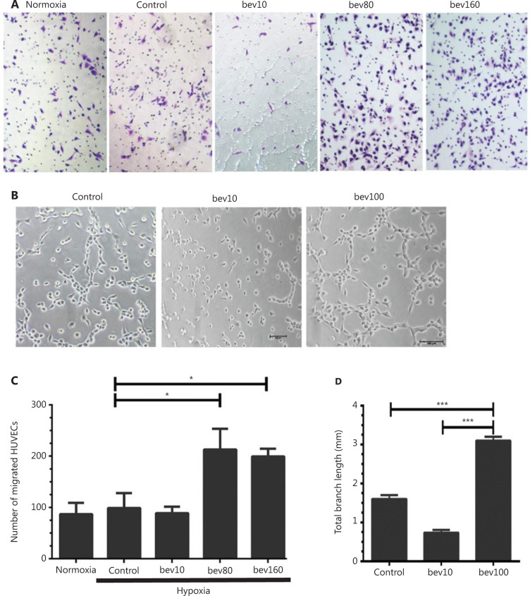
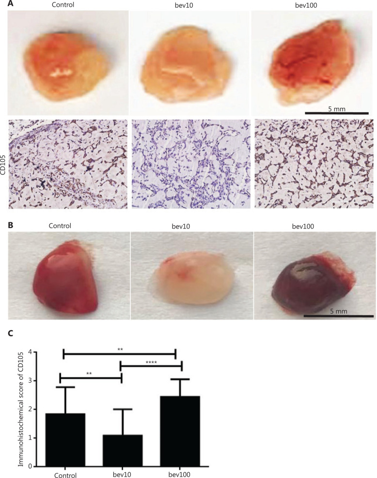
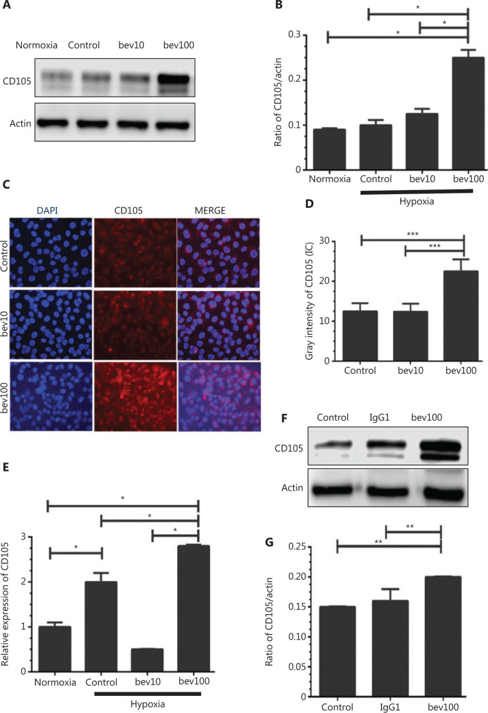
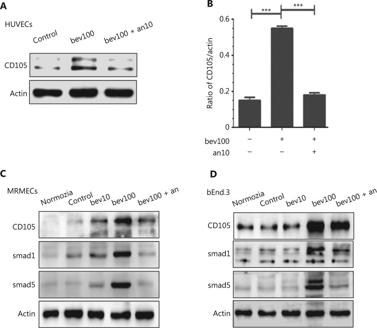
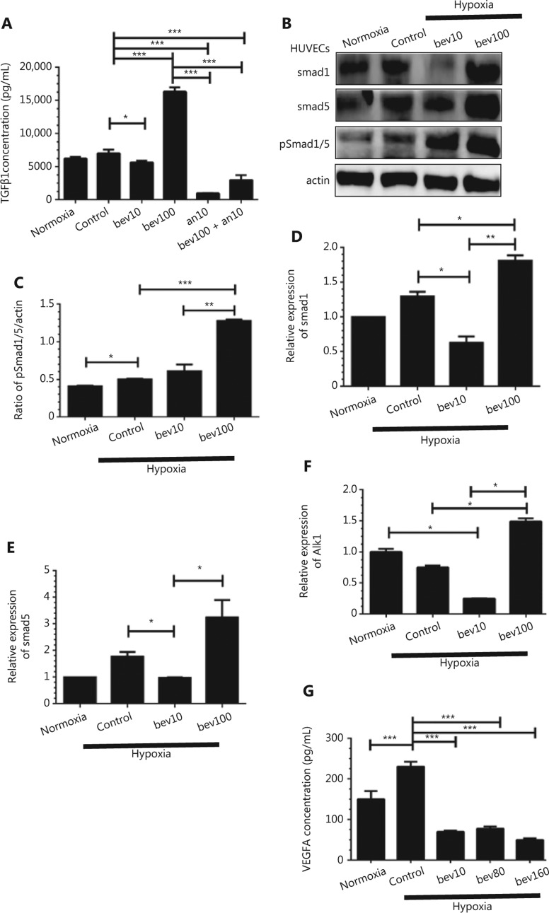
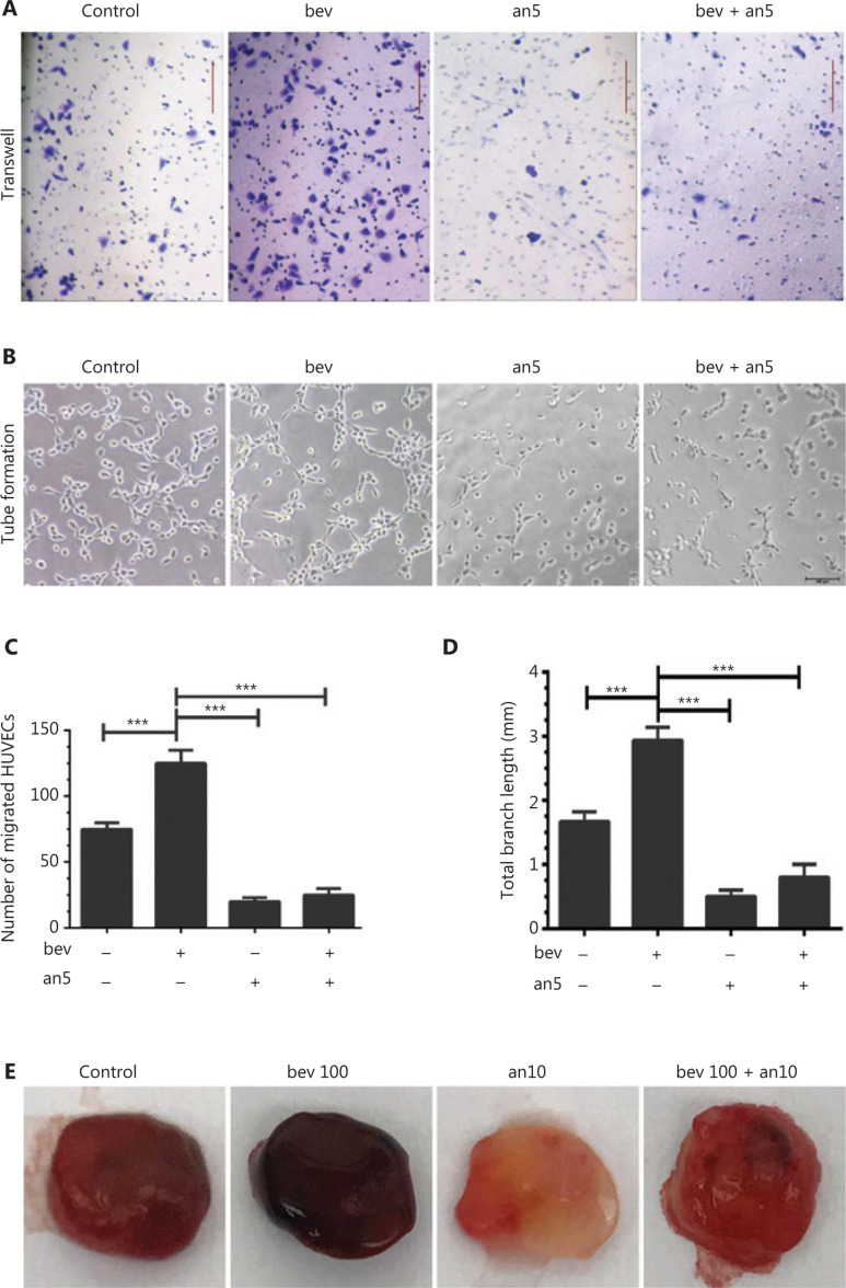
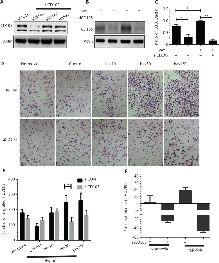
Objective: Bevacizumab is a recombinant humanized monoclonal antibody that blocks vascular endothelial growth factor (VEGF) with clear clinical benefits. However, overall survival of some cancer types remains low owing to resistance to bevacizumab therapy. While resistance is commonly ascribed to tumor cell invasion induced by hypoxia-inducible factor (HIF), less attention has been paid to the potential involvement of endothelial cells (ECs) in vasculature activated by anti-angiogenic drugs. Methods: Human umbilical vein ECs (HUVECs), bEnd.3 cells, and mouse retinal microvascular ECs (MRMECs) were treated with bevacizumab under conditions of hypoxia and effects on biological behaviors, such as migration and tube formation, examined. Regulatory effects on TGFβ1 and CD105 (endoglin) were established via determination of protein and mRNA levels. We further investigated whether the effects of bevacizumab could be reversed using the receptor tyrosine kinase inhibitor anlotinib. Results: Bevacizumab upregulated TGFβ1 as well as CD105, a component of the TGFβ receptor complex and an angiogenesis promoter. Elevated CD105 induced activation of Smad1/5, the inflammatory pathway and endothelial-mesenchymal transition. The migration ability of HUVECs was enhanced by bevacizumab under hypoxia. Upregulation of CD105 was abrogated by anlotinib, which targets multiple receptor tyrosine kinases including VEGFR2/3, FGFR1-4, PDGFRα/β, C-Kit, and RET. Conclusions: Bevacizumab promotes migration and tube formation of HUVECs via activation of the TGFβ1 pathway and upregulation of CD105 expression. Anlotinib reverses the effects of bevacizumab by inhibiting the above signals.

Keywords: CD105; HUVEC; TGFβ; anlotinib; bevacizumab.


Conflict of interest statement

*These authors contributed equally to this work.

Figures

Figure 1
Figure 1
High concentration of bevacizumab (100 μg/mL) enhances migration and tube formation of HUVECs in vitro. (A) Typical images of migrating HUVECs treated with bevacizumab under hypoxia conditions (hypoxia: O2 < 1%, 5% CO2, 94% N2; control: bevacizumab 0 μg/mL; bev10: bevacizumab 10 μg/mL, bev100: bevacizumab 100 μg/mL; normoxia: normal oxygen vehicle: 21% O2, 5% CO2, 74% N2); magnification, ×100. (B) Images of canal-like tubules formed by HUVECs treated with bevacizumab under hypoxia; magnification, ×50. (C) Quantitative analysis of migrating HUVECs treated with different doses of bevacizumab under hypoxia. Data represent mean ± SD of three independent experiments; *P < 0.05; one-way ANOVA. (D) Average total branching lengths of canal-like tubules formed by HUVECs treated with bevacizumab under hypoxia. Data represent mean ± SD, *P < 0.05; **P < 0.01; ***P < 0.001; one-way ANOVA.
Figure 2
Figure 2
High concentration of bevacizumab (100 μg/mL) accelerates angiogenesis of HUVECs and bEnd.3 in vivo. (A) Comparison of blood vessel formation in matrigel (400 μL) plugs in female nude mice (n = 4 per group) by bev (bevacizumab: 0 μg/mL, 10 μg/mL and 100 μg/mL in matrigel). Mixed matrigel containing HUVECs, bevacizumab, and VEGFA was subcutaneously injected into mice. Mice were intraperitoneally injected with 0, 5, and 50 mg/kg bevacizumab twice a week for 1 month. The image shows matrigel separated from mice, with darker red indicative of higher blood content in vasculature in the gel. CD105 expression (brown: CD105+, the antibody was only reactive to human endothelial cells) determined via immunochemical assay. The CD105+ stain was stronger in HUVECs treated with high concentrations of bevacizumab than those treated with low concentrations of bevacizumab. (B) Comparison of blood vessel formation in matrigel (400 μL) plugs in female nude mice (n = 4 per group) from control (bevacizumab: 0 μg/mL in matrigel), bev10 (bevacizumab: 10 μg/mL in matrigel), and bev100 (bevacizumab: 100 μg/mL in matrigel) groups. Mixed matrigel containing bEnd.3 cells, bevacizumab, and VEGFA was subcutaneously injected into mice, followed by intraperitoneal injection with 0, 5, or 50 mg/kg bevacizumab twice a week for 9 days. The image shows matrigels separated from mice, with darker red indicative of higher blood content in vasculature in the gel. (C) Histogram displaying immunochemistry scores of CD105 in matrigel containing HUVECs (n = 22 per group, data represent mean ± SD, **P < 0.01, ****P < 0.0001; non-parametric test).
Figure 3
Figure 3
High concentration of bevacizumab (100 μg/mL) stimulates CD105 expression. (A) Western blot showing changes in CD105 protein levels following bevacizumab treatment under normoxia and hypoxia conditions (control: bevacizumab 0 μg/mL, bev10: bevacizumab 10 μg/mL, bev100: bevacizumab 100 μg/mL; normoxia: normal oxygen vehicle). (B) Quantitative analysis of CD105 protein levels following bevacizumab treatment under hypoxia. Data represent mean ± SD, *P < 0.05; one-way ANOVA. (C) Immunofluorescence of CD105 in HUVECs pre-stimulated with bevacizumab under hypoxia (control: bevacizumab 0 μg/mL, bev10: bevacizumab 10 μg/mL, bev100: bevacizumab 100 μg/mL), CD105 (red), and DAPI (blue). Magnification, ×200. (D) Quantitative analysis of fluorescence intensity of CD105+, ***P < 0.001; one-way ANOVA. (E) Changes in CD105 mRNA levels in response to bevacizumab under hypoxia conditions. Data represent mean ± SD, *P < 0.05; one-way ANOVA. (F) Western blot showing CD105 expression upon treatment with 100 μg/mL bevacizumab and isotype control IgG1 under hypoxia. (G) Quantitative analysis of CD105 protein expression following bevacizumab (100 μg/mL) and isotype control (IgG1) treatment. Data represent mean ± SD, **P < 0.01; one-way ANOVA.
Figure 4
Figure 4
Bevacizumab upregulates CD105 and activates the TGFβ pathway in different cell lines under hypoxia, which is reversed by anlotinib regardless of the treatment sequence. (A) HUVECs were treated with bevacizumab (100 μg/mL, 24 h) or anlotinib (10 μM, 12 h) following pretreatment with bevacizumab (100 μg/mL, 12 h) under hypoxia. Anlotinib reversed bevacizumab-induced elevation of CD105. (B) Densitometric analysis of CD105 protein levels shown in (A). Data represent mean ± SD, ***P < 0.001, ANOVA. (C, D) MRMECs and bEnd.3 cells were treated with bevacizumab (0, 10, 100 μg/mL, 24 h) or anlotinib (10 μM, 12 h) following pretreatment with bevacizumab (100 μg/mL, 12 h) under hypoxia. Anlotinib reversed bevacizumab-induced elevation of CD105. Bevacizumab additionally enhanced Smad1 and Smad5 expression.
Figure 5
Figure 5
The TGFβ pathway is activated by high concentration of bevacizumab (100 μg/mL) under hypoxia conditions. (A) Concentrations of secreted TGFβ1 in supernatant determined using ELISA under hypoxia. HUVECs were treated with 10 and 100 μg/mL bevacizumab (24 h), anlotinib 10 μM (24 h), bevacizumab (100 μg/mL for 8 h) and anlotinib (10 μM for 16 h) sequentially; normoxia: normal oxygen vehicle. *P < 0.05; ***P < 0.001, one-way ANOVA. (B) Western blot showing changes in Smad1, Smad5, and pSmad1/5 protein levels (downstream factor of TGFβ and CD105) following bevacizumab treatment under hypoxia. (C) Quantitative analysis of pSmad1/5 protein levels following bevacizumab treatment. Data represent mean ± SD, *P < 0.05, **P < 0.01, ***P < 0.001; one-way ANOVA. (D–F) Quantitative analysis of Smad1, Smad5, and alk1 mRNA levels following treatment with bevacizumab under hypoxia, *P < 0.05, **P < 0.01, ***P < 0.001; one-way ANOVA. (G) Concentrations of secreted VEGFA in the supernatant under hypoxia, as determined with ELISA. HUVECs were treated with 10, 80, and 160 μg/mL bevacizumab (starvation for 12 h); normoxia: normal oxygen vehicle, *P < 0.05, **P < 0.01, ***P < 0.001; one-way ANOVA.
Figure 6
Figure 6
Anlotinib suppresses HUVEC migration and tube formation. (A) Typical images of HUVECs in migration assays following treatment with or without anlotinib and bevacizumab under hypoxia (an5: anlotinib 5 μM, bev: bevacizumab 100 μg/mL). Magnification, ×100. (B) Images of canal-like tubes formed by HUVECs treated with or without anlotinib and bevacizumab under hypoxia (an5: anlotinib 5 μM, bev: bevacizumab 100 μg/mL), Magnification, ×50. (C) Number of migrated HUVECs. Data represent mean ± SD, *P < 0.05, **P < 0.01, ***P < 0.001; one-way ANOVA. (D) Average total branching lengths of the capillary-like tubules formed following anlotinib and bevacizumab treatment under hypoxia. Data represent mean ± SD, *P < 0.05, **P < 0.01, ***P < 0.001; one-way ANOVA. (E) Comparison of blood vessel formation in matrigel (400 μL) plugs in female nude mice (n = 4 per group) between control (bevacizumab: 0 μg/mL in matrigel), bev100 (bevacizumab: 100 μg/mL in matrigel) and bev100 + an10 (100 μg/mL bevacizumab and 10 μM anlotinib in matrigel) groups. Blood and vessel structures were evident in the bev100 and control groups, but bEnd.3 cells treated with bevacizumab and anlotinib displayed no obvious vessel structures in the matrigel. Mixed matrigel containing bEnd.3 cells, bevacizumab, and VEGFA was subcutaneously injected into mice, followed by intraperitoneal injection with 0, 5, and 50 mg/kg bevacizumab twice a week for 9 days. The image shows bEnd.3 matrigel separated from mice, with darker red indicative of higher blood content in the gel.
Figure 7
Figure 7
CD105 siRNA suppresses CD105 expression as well as migration and proliferation of HUVECs promoted by bevacizumab. (A) CD105 protein levels following siCD105 (CD105 siRNA) treatment determined via Western blot. The CD105 siRNA sequences are as follows: siRNA1, 5′-CCA UGA CCC UGG UAC UAA A-3′ and 3′-GGU ACU GGG ACC AUG AUU U-5′; siRNA2, 5′-UGA CCU GUC UGG UUG CAC ATT-3′ and 5′-UGU GCA ACC AGA CAG GUC AGG-3′; siRNA3, 5′-GAG GUG ACA UAU ACC ACU A-3′, 5′-CUC CAC UGU AUA UGG UGA U-3′; and siCON, 5′-UUC UCC GAA CGU GUC ACG UTT-3′ and 5′-ACG UGA CAC GUU CGGAGAATT-3′. CD105 was markedly downregulated by siRNA1, which was selected for subsequent experiments. (B) Western blot assessment of CD105 protein levels following bevacizumab treatment in the presence or absence of siCD105 (CD105 siRNA). (C) Densitometry analysis of CD105 protein levels from B. Data represent mean ± SD, *P < 0.05, **P < 0.01; Student’s t-test. (D) Typical images of migrated HUVECs in transwell assays following bevacizumab treatment in the presence or absence of CD105 siRNA (siCD105); normoxia: normal oxygen vehicle, control: hypoxia, bevacizumab 0 μg/mL, bev10, bev80, bev160: bevacizumab 10 μg/mL, 80 μg/mL, 160 μg/mL. (E) Number of migrated HUVECs. Data represent mean ± SD, **P < 0.01; Student’s t-test. (F) Proliferation rate of HUVECs decreased following treatment with siRNAs targeting CD105 under both normal and hypoxia conditions. Data represent mean ± SD.

References

    1. Chu BF, Otterson GA. Incorporation of antiangiogenic therapy into the non-small-cell lung cancer paradigm. Clin Lung Cancer. 2016;17:493–506. - PubMed
    1. Hurwitz H, Fehrenbacher L, Novotny W, Cartwright T, Hainsworth J, Heim W, et al. Bevacizumab plus irinotecan, fluorouracil, and leucovorin for metastatic colorectal cancer. N Engl J Med. 2004;350:2335–42. - PubMed
    1. Reck M, von Pawel J, Zatloukal P, Ramlau R, Gorbounova V, Hirsh V, et al. Overall survival with cisplatin-gemcitabine and bevacizumab or placebo as first-line therapy for nonsquamous non-small-cell lung cancer: results from a randomised phase III trial (AVAiL). Ann Oncol. 2010;21:1804–9. - PMC - PubMed
    1. Robert NJ, Dieras V, Glaspy J, Brufsky AM, Bondarenko I, Lipatov ON, et al. RIBBON-1: randomized, double-blind, placebo-controlled, phase III trial of chemotherapy with or without bevacizumab for first-line treatment of human epidermal growth factor receptor 2-negative, locally recurrent or metastatic breast cancer. J Clin Oncol. 2011;29:1252–60. - PubMed
    1. Saltz LB, Clarke S, Diaz-Rubio E, Scheithauer W, Figer A, Wong R, et al. Bevacizumab in combination with oxaliplatin-based chemotherapy as first-line therapy in metastatic colorectal cancer: a randomized phase III study. J Clin Oncol. 2008;26:2013–9. - PubMed

Publication types

MeSH terms

LinkOut - more resources