. 2020 Jun 16:8:e9298.
doi: 10.7717/peerj.9298. eCollection 2020.

Combination of ferric ammonium citrate with cytokines involved in apoptosis and insulin secretion of human pancreatic beta cells related to diabetes in thalassemia

Affiliations

Combination of ferric ammonium citrate with cytokines involved in apoptosis and insulin secretion of human pancreatic beta cells related to diabetes in thalassemia

Patchara Rattanaporn et al. PeerJ. .

Abstract

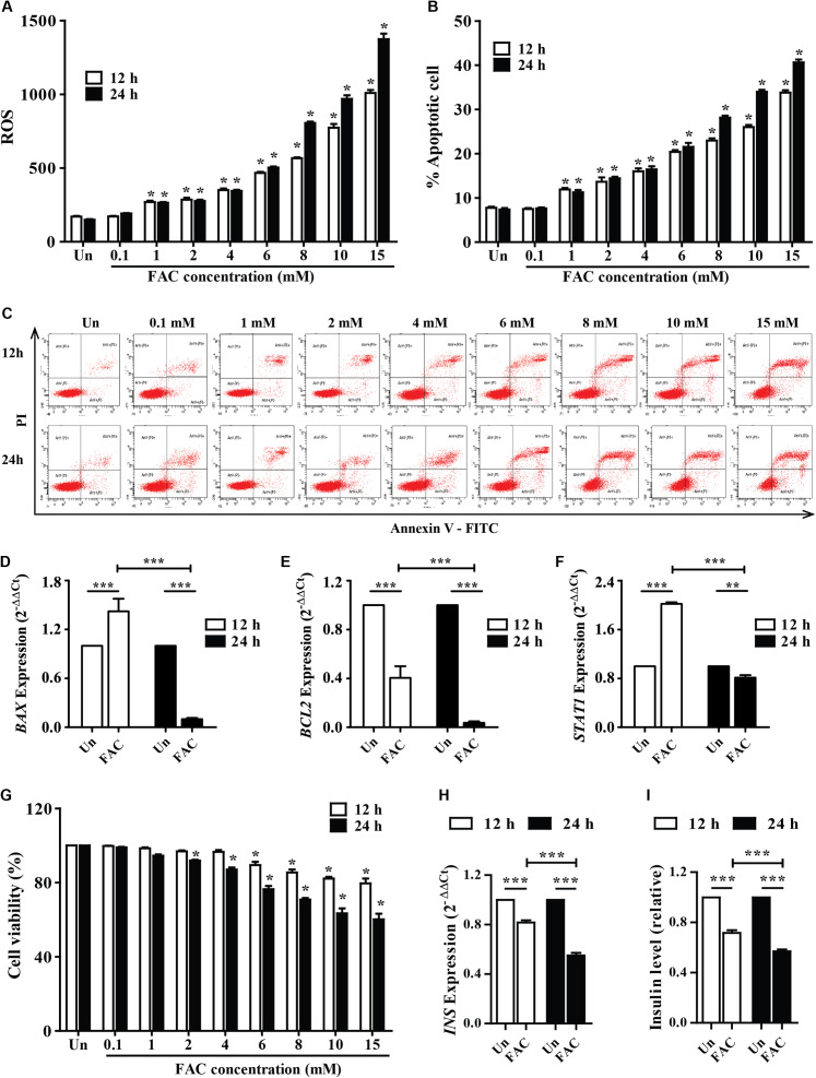
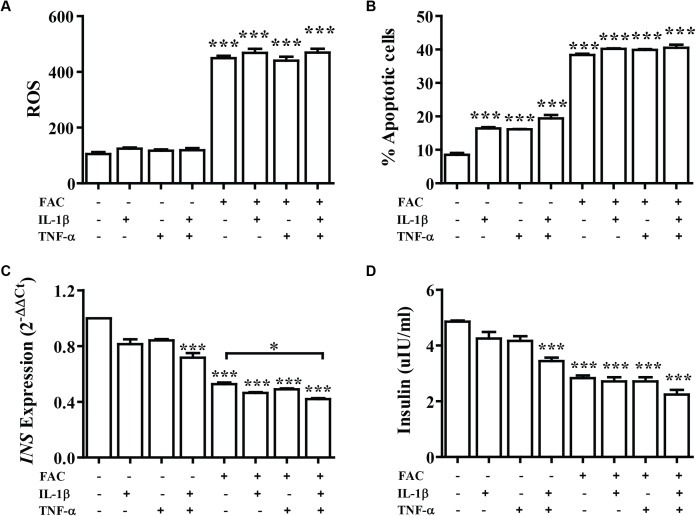
Background: Diabetes mellitus (DM) is a common complication found in β-thalassemia patients. The mechanism of DM in β-thalassemia patients is still unclear, but it could be from an iron overload and increase of some cytokines, such as interleukin1-β (IL-1β) and tumor necrosis factor-α (TNF-α). The objective of this study was to study the effect of interaction between ferric ammonium citrate (FAC) and cytokines, IL-1β and TNF-α, on 1.1B4 human pancreatic β-cell line.

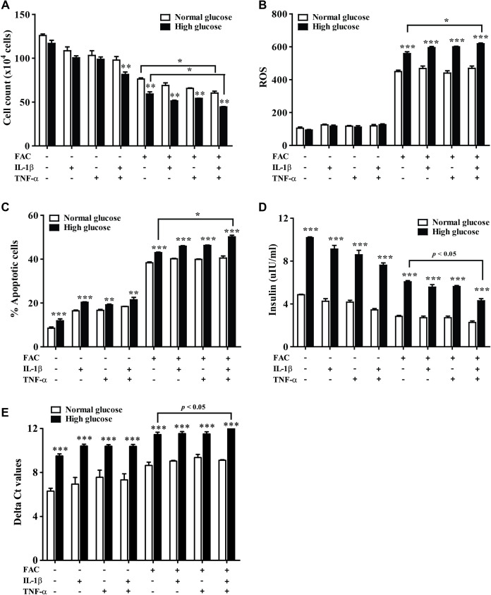
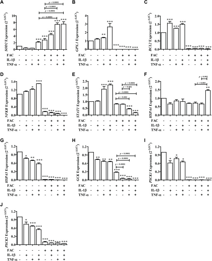
Methods: The effect of the combination of FAC and cytokines on cell viability was studied by MTT assay. Insulin secretion was assessed by the enzyme-linked immunosorbent assay (ELISA). The reactive oxygen species (ROS) and cell apoptosis in normal and high glucose condition were determined by flow cytometer. In addition, gene expression of apoptosis, antioxidant; glutathione peroxidase 1 (GPX1) and superoxide dismutase 2 (SOD2), and insulin secretory function were studied by real-time polymerase chain reaction (Real-time PCR).

Results: The findings revealed that FAC exposure resulted in the decrease of cell viability and insulin-release, and the induction of ROS and apoptosis in pancreatic cells. Interestingly, a combination of FAC and cytokines had an additive effect on SOD2 antioxidants' genes expression and endoplasmic reticulum (ER) stress. In addition, it reduced the insulin secretion genes expression; insulin (INS), glucose kinase (GCK), protein convertase 1 (PSCK1), and protein convertase 2 (PSCK2). Moreover, the highest ROS and the lowest insulin secretion were found in FAC combined with IL-1β and TNF-α in the high-glucose condition of human pancreatic beta cell, which could be involved in the mechanism of DM development in β-thalassemia patients.

Keywords: Apoptosis; Cytokines; Diabetes; ER stress; Ferric ammonium citrate; Insulin; Iron overload; Pancreatic beta cell; Reactive oxygen species; Thalassemia.


Conflict of interest statement

The authors declare that they have no competing interests.

Figures

Figure 1
Figure 1. Iron-induce intracellular ROS generation, increase cell death, reduce INS expression, and insulin production in pancreatic β-cells.
The 1.1B4 cells were exposed to different concentrations of FAC for 12 and 24 h. (A) ROS, cells stained with DCFH-DA were detected using a flow cytometer. (B) Apoptosis, cells were stained with fluorescein isothiocyanate (FITC)-conjugated Annexin V and PI and was detected by using a flow cytometer. (C) Flow plots of apoptosis. Apoptosis regulatory gene expression, mRNA were extracted and converted to cDNA. The mRNA expression was normalized to ACTB expression; (D) BAX expression. (E) BCL2 expression. (F) STAT1: expression. (G) cell viability was measured using MTT assay. The INS expression was checked, and the cultured medium was collected for insulin measurement using an ELISA kit. (H) INS expression in 2−ΔΔCt. Later, it was normalized by ACTB. (I) insulin level, which was normalized by the untreated cells. The results were shown in mean ± SEM from three independent experiments and yielded. *p < 0.05 and ** p < 0.001 indicate a statistically difference compared to the untreated cells. ***p < 0.0001, indicating a statistically significant difference compared with untreated cells and 12 h using a one-way ANOVA test.
Figure 2
Figure 2. The effect of FAC combine with IL-1β and TNF-α.
The 1.1B4 cells were treated with 8 mM FAC combined with 10 pg/mL of IL-1β, FAC combined with 20 pg/mL of TNF-α, and FAC combined with IL-1β and TNF-α for 24 h. (A) ROS cells stained with DCFH-DA were detected using a flow cytometer. (B) Apoptosis cells stained with fluorescein isothiocyanate (FITC)-conjugated AnV and PI were detected using a flow cytometer. (C) INS expression, mRNA were extracted and converted to cDNA. The mRNA expression was normalized to ACTB expression. (D) Insulin level. The culture medium was collected for insulin measurement using the ELISA technique. The results are shown in mean ± SEM from three independent experiments and yield a ***p < 0.0001, indicating a statistically significant difference compared with the untreated cells and a *p < 0.05, indicating a statistically significant difference compared with FAC treated alone.
Figure 3
Figure 3. The effect of high glucose on iron combined with IL-1β- and TNF-α-treated cells.
The 1.1B4 cells were treated with 8 mM FAC combined with 10 pg/mL of IL-1β, FAC combined with 20 pg/mL of TNF-α, and FAC combined with IL-1β and TNF-α in no adding glucose and 22.2 mM glucose adding condition for 24 h. (A) Cell count, the cells stained with typhan blue were counted under a microscope. (B) The ROS, cells stained with DCFH-DA were detected using a flow cytometer. (C) The apoptosis cells stained with fluorescein isothiocyanate (FITC)-conjugated AnV and PI were detected using a flow cytometer. (D) Insulin level, the culture medium was collected for insulin measurement using the ELISA technique. (E) INS expression, mRNA were extracted and converted to cDNA. The mRNA expression was shown as delta Ct values calculated from CtINS to CtACTB. The results are shown in mean ± SEM of three independent experiments and revealed a **p < 0.01 and a ***p < 0.0001, indicating the statistically significant differences compared to the no adding glucose. *represent the significantly difference at p < 0.05.
Figure 4
Figure 4. The expression of genes involved in insulin secretion, antioxidants, ER stress, and apoptosis signaling pathways.
Following FAC and cytokines exposure, mRNA was extracted and converted to cDNA. The mRNA expression was normalized to ACTB expression. The results are shown as mean ± SEM of three independent experiments and indicated a *p < 0.05, **p < 0.01, and ***p < 0.0001, indicating the statistically significant differences compared to the untreated cells. The antioxidant genes include SOD2 (A) and GPX.1 (B). The apoptosis pathway genes are BCL2 (C), NFκB (D), and STAT1 (E). ER stress genes include HSPA4 (F) and HSPA5 (G). The secretory function genes include GCK (H), PSCK1 (I), and PSCK2 (J).

Similar articles

Cited by

References

    1. Andrews NC. The iron transporter DMT1. International Journal of Biochemistry and Cell Biology. 1999;31(10):991–994. doi: 10.1016/S1357-2725(99)00065-5. - DOI - PubMed
    1. Berchtold LA, Prause M, Storling J, Mandrup-Poulsen T. Cytokines and pancreatic beta-cell apoptosis. Advances in Clinical Chemistry. 2016;75:99–158. - PubMed
    1. Bogdan AR, Miyazawa M, Hashimoto K, Tsuji Y. Regulators of iron homeostasis: new players in metabolism, cell death, and disease. Trends in Biochemical Sciences. 2016;41(3):274–286. doi: 10.1016/j.tibs.2015.11.012. - DOI - PMC - PubMed
    1. Cao JY, Dixon SJ. Mechanisms of ferroptosis. Cellular and Molecular Life Sciences. 2016;73(11–12):2195–2209. doi: 10.1007/s00018-016-2194-1. - DOI - PMC - PubMed
    1. Dixon SJ, Stockwell BR. The role of iron and reactive oxygen species in cell death. Nature Chemical Biology. 2014;10(1):9–17. doi: 10.1038/nchembio.1416. - DOI - PubMed

LinkOut - more resources