Proliferation, Characterization and Differentiation Potency of Adipose Tissue-Derived Mesenchymal Stem Cells (AT-MSCs) Cultured in Fresh Frozen and non-Fresh Frozen Plasma
- PMID: 32587838
- PMCID: PMC7305462
- DOI: 10.22088/IJMCM.BUMS.8.4.283
Proliferation, Characterization and Differentiation Potency of Adipose Tissue-Derived Mesenchymal Stem Cells (AT-MSCs) Cultured in Fresh Frozen and non-Fresh Frozen Plasma
Abstract
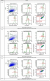
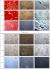
Mesenchymal stem cells (MSCs) have unique properties, including high proliferation rates, self-renewal, and multilineage differentiation ability. Their characteristics are affected by increasing age and microenvironment. This research is aimed to determine the proliferation, characteristics and differentiation capacity of adipose tissue-derived (AT)-MSCs at many passages with different media. The cell proliferation capacity was assayed using trypan blue. MSCs characterization (CD90, CD44, CD105, CD73, CD11b, CD19, CD34, CD45, and HLA-DR) was performed by flow cytometry, and cell differentiation was determined by specific stainings. Population doubling time (PDT) of AT-MSCs treated with fresh frozen plasma (FFP) and non-FFP increased in the late passage (P) (P15 FFP was 22.67 ± 7.01 days and non-FFP was 19.65 ± 2.27 days). Cumulative cell number was significantly different between FFP and non-FFP at P5, 10, 15. AT-MSCs at P4-15 were positive for CD90, CD44, CD105, and CD73, and negative for CD11b, CD19, CD34, CD45, and HLA-DR surface markers. AT-MSCs at P5, 10, 15 had potential toward adipogenic, chondrogenic, and osteogenic differentiation. Therefore, PDT was affected by increased age but no difference was observed in morphology, surface markers and differentiation capacity among passages. Cumulative cell number in FFP was higher in comparison with non-FFP in P5, 10, 15. Our data suggest that FFP may replace FBS for culturing MSCs.
Keywords: Adipose tissue-MSCs; multilineage differentiation; population doubling time; proliferation; surface marker.
Figures


References
-
- Widowati W, Wijaya L, Murti H, et al. Conditioned medium from normoxia (WJMSCs-norCM) and hypoxia-treated WJMSCs (WJMSCs-hypoCM) in inhibiting cancer cell proliferation. Biomarkers Genomic Med. 2015;7:8–17.
-
- Widowati W, Murti H, Jasaputra DK, et al. Selective cytotoxic potential of IFN-γ and TNF-α on breast cancer cell lines (T47D and MCF7) Asian J Cell Biol. 2016;11:1–12.
-
- Widowati W, Wijaya L, Bachtiar I, et al. Effect of oxygen tension on proliferation and characteristics of Wharton's jelly-derived mesenchymal stem cells. Biomarkers Genomic Med. 2014;6:43–8.
-
- Widowati W, Krisanti Jasaputra D, B Sumitro S, et al. Potential of Unengineered and Engineered Wharton’s Jelly Mesenchymal Stem Cells as Cancer Inhibitor Agent. Immun Endor Metab Agents in Med Chem. 2015;15:128–37.
LinkOut - more resources
Full Text Sources
Research Materials
Miscellaneous
