. 2020 May 25;4(7):bvaa060.
doi: 10.1210/jendso/bvaa060. eCollection 2020 Jul 1.

Recurrent Amplification of the Osmotic Stress Transcription Factor NFAT5 in Adrenocortical Carcinoma

Affiliations

Recurrent Amplification of the Osmotic Stress Transcription Factor NFAT5 in Adrenocortical Carcinoma

Taylor C Brown et al. J Endocr Soc. .

Abstract

Tumorigenesis requires mitigation of osmotic stress and the transcription factor nuclear factor of activated T cells 5 (NFAT5) coordinates this response by inducing transcellular transport of ions and osmolytes. NFAT5 modulates in vitro behavior in several cancer types, but a potential role of NFAT5 in adrenocortical carcinoma (ACC) has not been studied. A discovery cohort of 28 ACCs was selected for analysis. Coverage depth analysis of whole-exome sequencing reads assessed NFAT5 copy number alterations in 19 ACCs. Quantitative real-time PCR measured NFAT5 mRNA expression levels in 11 ACCs and 23 adrenocortical adenomas. Immunohistochemistry investigated protein expression in representative adrenal samples. The Cancer Genome Atlas database was analyzed to corroborate NFAT5 findings from the discovery cohort and to test whether NFAT5 expression correlated with ion/osmolyte channel and regulatory protein expression patterns in ACC. NFAT5 was amplified in 10 ACCs (52.6%) and clustered in the top 6% of all amplified genes. mRNA expression levels were 5-fold higher compared with adrenocortical adenomas (P < 0.0001) and NFAT5 overexpression had a sensitivity and specificity of 81.8% and 82.7%, respectively, for malignancy. Increased protein expression and nuclear localization occurred in representative ACCs. The Cancer Genome Atlas analysis demonstrated concomitant NFAT5 amplification and overexpression (P < 0.0001) that correlated with increased expression of sodium/myo-inositol transporter SLC5A3 (r 2 = 0.237, P < 0.0001) and 14 other regulatory proteins (P < 0.05) previously shown to interact with NFAT5. Amplification and overexpression of NFAT5 and associated osmotic stress response related genes may play an important role adrenocortical tumorigenesis.

Keywords: Adrenocortical carcinoma; NFAT5; Osmotic Stress; and SLC5A3.


Figures

Figure 1.
Figure 1.
NFAT5 gene copy analysis. (A) Nineteen samples previously underwent WES (15). Gene copy number was determined by assessing the ratio of coverage depth of WES reads between tumor and adjacent normal adrenal DNA. The NFAT5 gene was significantly affected, ranking in the top 6% of all amplified genes. Overall, NFAT5 gene copy alterations significantly deviated from normal diploid copy number (*P = 0.0068). (B) Analysis of the TCGA ACC cohort database demonstrated similar gene copy alterations in the NFAT5 locus (*P < 0.0001). NFAT5, nuclear factor of activated T cells 5; TCGA, The Cancer Genome Atlas; WES, whole-exome sequencing. Horizontal bar, mean; error bars, standard deviation.
Figure 2.
Figure 2.
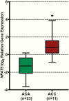
NFAT5 gene expression analysis. Relative messenger RNA expression levels of NFAT5 in ACCs (n = 11) were measured by real-time quantitative PCR and compared with expression levels in ACAs (n = 23). Twelve samples of normal adrenal tissue served as a reference control. Overall expression levels were approximately 5-fold higher in ACC compared with ACA (*P < 0.0001). ACA, adrenocortical adenoma; ACC, adrenocortical carcinoma; NFAT5, nuclear factor of activated T cells 5. Horizontal bar, median; error bars, minimum and maximum.
Figure 3.
Figure 3.
NFAT5 immunohistochemical analysis. Immunostaining of NFAT5 in a representative sample of ACC compared with ACA and normal adrenal tissue. Overall, protein expression levels were higher with increased expression in ACC compared with ACA and normal samples. Furthermore, increased nuclear localization of NFAT5 protein was predominantly observed in ACC samples. Magnification, 100× and 400×; NFAT5 = brown. ACA, adrenocortical adenoma; ACC, adrenocortical carcinoma; NFAT5, nuclear factor of activated T cells 5.
Figure 4.
Figure 4.
NFAT5 gene copy gains and expression. When tumors were stratified by NFAT5 gene copy status (deletions, diploid, amplifications), gene expression levels were tightly correlated with gene copy number (*P < 0.0001). NFAT5, nuclear factor of activated T cells 5. Horizontal bar, mean; error bars, standard deviation.
Figure 5.
Figure 5.
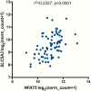
NFAT5 and SLC5A3 expression. Messenger RNA expression of SLC5A3 was associated with NFAT5 mRNA expression (r2 = 0.237, P < 0.0001). NFAT5, nuclear factor of activated T cells 5; SLC5A3, solute carrier family 5 member 3.
Figure 6.
Figure 6.
NFAT5 signaling in adrenocortical carcinoma. (A) Messenger RNA expression levels of 5 representative proteins previously shown to interact with and/or regulate NFAT5 were significantly correlated with NFAT5 expressions levels (P < 0.05). (B) STRING (version 11.0) analysis revealed a statistically significant association of 14 proteins shown in the TCGA cohort to be coexpressed with NFAT5. Black arrows mark genes highly correlated with NFAT5 expression.

Similar articles

Cited by

References

    1. Sharma E, Dahal S, Sharma P, et al. . The characteristics and trends in adrenocortical carcinoma: a United States Population Based Study. J Clin Med Res. 2018;10(8):636-640. - PMC - PubMed
    1. Lebastchi AH, Kunstman JW, Carling T. Adrenocortical carcinoma: current therapeutic state-of-the-art. J Oncol. 2012;2012:234726. - PMC - PubMed
    1. Amini N, Margonis GA, Kim Y, et al. . Curative resection of adrenocortical carcinoma: rates and patterns of postoperative recurrence. Ann Surg Oncol. 2016;23(1):126-133. - PMC - PubMed
    1. Tella SH, Kommalapati A, Yaturu S, Kebebew E. Predictors of survival in adrenocortical carcinoma: an analysis from the national cancer database. J Clin Endocrinol Metab. 2018;103(9):3566-3573. - PubMed
    1. Megerle F, Kroiss M, Hahner S, Fassnacht M. Advanced adrenocortical carcinoma - what to do when first-line therapy fails? Exp Clin Endocrinol Diabetes. 2019;127(2-03):109-116. - PubMed

LinkOut - more resources