

Role of diffusion-weighted imaging in the diagnosis of cerebral venous thrombosis
- PMID: 32589072
- PMCID: PMC7323280
- DOI: 10.1177/0300060520933448
Role of diffusion-weighted imaging in the diagnosis of cerebral venous thrombosis
Abstract
Objective: To evaluate the hyperintense signal (HIS) performance on diffusion-weighted imaging (DWI) in diagnosing cerebral venous thrombosis (CVT).
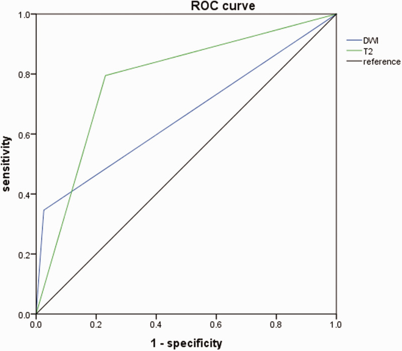
Methods: Seventy-eight patients with CVT hospitalized from January 2004 to January 2015 were retrospectively studied alongside 78 controls without intracranial organic diseases. Diagnostic accuracy indices of HIS on DWI or T2-weighted imaging (T2WI) to diagnose CVT at different sites and states were analyzed.
Results: The overall sensitivity of HIS on DWI for the diagnosis of CVT was significantly lower than that of HIS on T2WI (34.6% vs. 79.5%). HIS on T2WI was more sensitive than HIS on DWI in detecting thrombosis, especially in the superior sagittal sinus and transverse sinus. HIS on DWI was inversely related to the time between disease onset and imaging. Compared with HIS on T2WI, combining HIS on DWI and T2WI did not increase the sensitivity for detecting CVT. HIS on DWI was not detected in the control group, but HIS on T2WI was detected in 26.3% of control individuals. The specificity of HIS on DWI for CVT was higher than that of HIS on T2WI (97.4% vs. 76.9%).
Conclusion: HIS on DWI has a lower sensitivity, but a higher specificity, than HIS on T2WI for diagnosing CVT.
Keywords: Cerebral venous thrombosis; T2-weighted imaging; diagnostic sensitivity; diagnostic specificity; diffusion-weighted imaging; hyperintense signal; magnetic resonance imaging.
Figures


References
-
- Stam J. Thrombosis of the cerebral veins and sinuses. N Eng J Med 2005; 353: 314–315. - PubMed
-
- Saposnik G, Barinagarrementeria F, Brown RD, et al. Diagnosis and management of cerebral venous thrombosis a statement for healthcare professionals from the American Heart Association/American Stroke Association. Stroke 2011; 42: 1158. - PubMed
-
- Corvol JC, Oppenheim C, Manai R, et al. Diffusion-weighted magnetic resonance imaging in a case of cerebral venous thrombosis. Stroke 1998; 29: 2649–2652. - PubMed
MeSH terms
LinkOut - more resources
Full Text Sources
Medical
