Comparative Study
. 2020 Aug 1;319(2):H331-H340.
doi: 10.1152/ajpheart.00274.2020. Epub 2020 Jun 26.

Pressure overload generates a cardiac-specific profile of inflammatory mediators

Affiliations
Comparative Study

Pressure overload generates a cardiac-specific profile of inflammatory mediators

Matthew O'Brien et al. Am J Physiol Heart Circ Physiol. .

Abstract

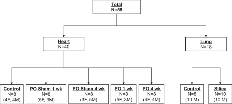
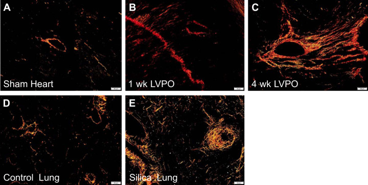
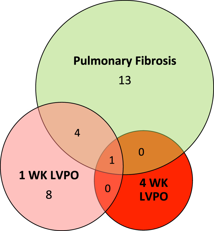
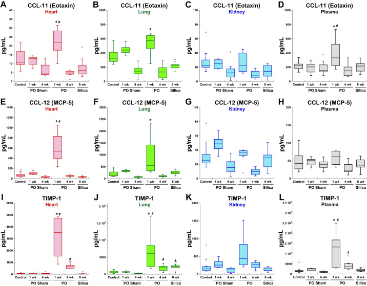
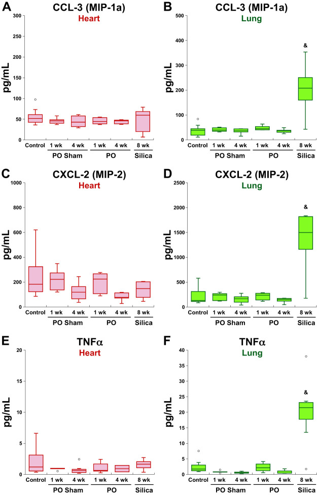
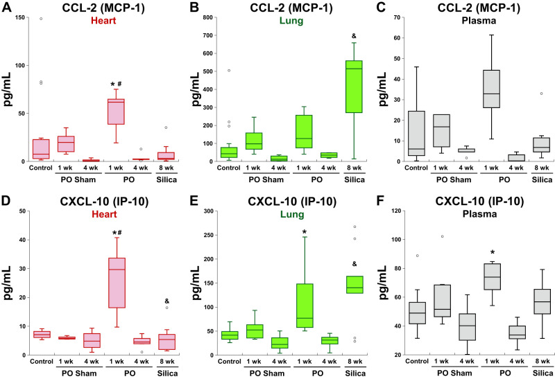
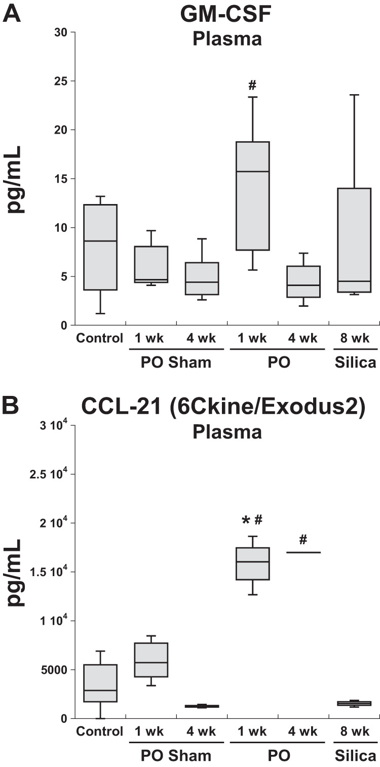
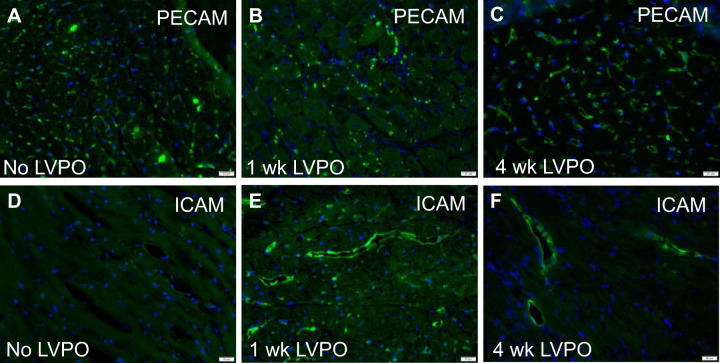
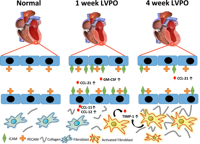
Mechanisms that contribute to myocardial fibrosis, particularly in response to left ventricular pressure overload (LVPO), remain poorly defined. To test the hypothesis that a myocardial-specific profile of secreted factors is produced in response to PO, levels of 44 factors implicated in immune cell recruitment and function were assessed in a murine model of cardiac hypertrophy and compared with levels produced in a model of pulmonary fibrosis (PF). Mice subjected to PO were assessed at 1 and 4 wk. Protein from plasma, LV, lungs, and kidneys were analyzed by specific protein array analysis in parallel with protein from mice subjected to silica-instilled PF. Of the 44 factors assessed, 13 proteins were elevated in 1-wk PO myocardium, whereas 18 proteins were found increased in fibrotic lung. Eight of those increased in 1-wk LVPO were not found to be increased in fibrotic lungs (CCL-11, CCL-12, CCL-17, CCL-19, CCL-21, CCL-22, IL-16, and VEGF). Additionally, six factors were increased in plasma of 1-wk LVPO in the absence of increases in myocardial levels. In contrast, in mice with PF, no factors were found increased in plasma that were not elevated in lung tissue. Of those factors increased at 1 wk, only TIMP-1 remained elevated at 4 wk of LVPO. Immunohistochemistry of myocardial vasculature at 1 and 4 wk revealed similar amounts of total vasculature; however, evidence of activated endothelium was observed at 1 wk and, to a lesser extent, at 4 wk LVPO. In conclusion, PO myocardium generated a unique signature of cytokine expression versus that of fibrotic lung.NEW & NOTEWORTHY Myocardial fibrosis and the resultant increases in myocardial stiffness represent pivotal consequences of chronic pressure overload (PO). In this study, cytokine profiles produced in a murine model of cardiac fibrosis induced by PO were compared with those produced in response to silica-induced lung fibrosis. A unique profile of cardiac tissue-specific and plasma-derived factors generated in response to PO are reported.

Keywords: cytokine; fibrosis; heart; inflammation; pressure overload; pulmonary.


Conflict of interest statement

No conflicts of interest, financial or otherwise, are declared by the authors.

Figures

Fig. 1.
Fig. 1.
Consort diagram describing the animal groups used in the present study. Forty mice were used in the cardiac pressure-overload (PO) arm of the experiment, whereas 18 animals were used for the silica-induced lung fibrosis studies. Both male (M) and female (F) mice were used in each arm.
Fig. 2.
Fig. 2.
Picrosirius red (PSR)-stained images of tissue sections taken from left ventricle of sham (A), 1-wk left ventricular pressure overload (LVPO; B), and 4-wk LVPO (C), demonstrating increases in histochemical staining of collagen with LVPO. In the silica-induced lung fibrosis model, sections of lung from 8-wk silica mice (E) had robust staining of fibrillar collagen vs. control (D). Images are representative of n = 5 mice (n = 3 sections/mouse). Size bar, 50 μm.
Fig. 3.
Fig. 3.
Venn diagram illustrating differences in cytokine/factor expression found in involved tissue in 1-wk left ventricular pressure overload (LVPO) myocardium (pink circle), silica-induced fibrotic lung (green circle), and 4-wk LVPO myocardium (red circle). For a complete list of cytokine/factors assessed, see Supplemental Table S1.
Fig. 4.
Fig. 4.
Representative factors elevated in left ventricular pressure overload (LVPO) myocardium. CC ligand (CCL)-11 (AD) and CCL-12 (EH) were increased in hearts and in lungs of 1 wk LVPO mice. No increases in CCL-11 or CCL-12 were found in fibrotic lung or in hearts or kidneys from silica-treated mice. Tissue inhibitor of matrix metalloproteinase-1 (TIMP-1; IL) was found to be increased in both LVPO myocardium and in lung as well as in silica-treated lung. TIMP-1 was the only factor assayed found to be significantly elevated in 4 wk LVPO myocardium, albeit in lower amounts than that of 1-wk LVPO. Plasma concentrations of of TIMP-1 were found to be elevated at both 1 and 4 wk of LVPO. Levels of CCL-11 were also found to be elevated in plasma from LVPO mice at 1 wk only, whereas plasma levels of CCL-12 were not increased in any of the tested mice. *P < 0.05 LVPO vs. control; #P < 0.05 LVPO vs. corresponding sham; &P < 0.05 silica vs. control. MCP-5, monocyte chemoattractant protein-5; PO, pressure overload.
Fig. 5.
Fig. 5.
Representative factors increased in lungs from silica-instilled mice that were not significantly elevated in fibrotic hearts from 1-wk or 4-wk left ventricular pressure overload (LVPO). CC ligand-3 (CCL-3; A and B), CXC ligand-2 (CXCL-2; C and D), and TNFα (E and F) were increased in fibrotic lungs but not induced in left ventricle (LV) at 1 wk or 4 wk of LVPO. Levels of CXCL-2 were also slightly elevated in the LV of silica-instilled mice. TNFα was found to slightly increase in the lungs of 1-wk LVPO, but not to the extent seen in fibrotic lung. CCL-3, but not CXCL-2 or TNFα, was significantly elevated in plasma from silica-instilled mice (data not shown). &P < 0.05 silica vs. control. MIP-1a and -2, macrophage inflammatory protein-1a and -2, respectively; PO, pressure overload.
Fig. 6.
Fig. 6.
Representative factors significantly elevated in fibrotic left ventricle (LV) from 1-wk left ventricular pressure overload (LVPO) myocardium and also increased in fibrotic lung. AC: levels of CC ligand-2 (CCL-2) were found to be elevated in lungs of 1-wk LVPO and plasma, whereas levels were not increased in hearts from silica-instilled mice nor in plasma. DF: levels of CXC ligand-10 (CXCL-10) were increased in LV, lungs, and plasma in 1-wk LVPO mice and in hearts from silica-instilled mice but were not found elevated in plasma from silica-instilled mice. *P < 0.05 LVPO vs. control; #P < 0.05 LVPO vs. corresponding sham; &P < 0.05 silica vs. control.
Fig. 7.
Fig. 7.
Representative factors found to be significantly elevated in plasma of 1-wk wk left ventricular pressure overload (LVPO) mice that were not elevated in plasma of mice with silica-instilled pulmonary fibrosis (PF). CC ligand-21 (CCL-21) remained elevated in plasma of mice with 4 wk of pressure overlaod (PO). Although CCL-21 was also elevated in 1 wk LVPO myocardium (B), granulocyte macrophage-colony stimulating factor (GM-CSF) was not increased in hearts at 1 wk of LVPO (data not shown; A). *P < 0.05, LVPO vs. control; #P < 0.05, LVPO vs. corresponding sham.
Fig. 8.
Fig. 8.
Immunohistochemical staining of vasculature in control (A), 1-wk (B), and 4-wk (C) left ventricular pressure overload (LVPO) using anti-platelet endothelial cell adhesion molecule (PECAM) PECAM antibodies revealed no overt differences in total vessel density or size after LVPO (quantification in Supplemental Material). Intercellular adhesion molecule (ICAM) immunoreactivity, indicative of activated endothelium, was visibly increased at 1 wk of LVPO (E) over control (D) with diminished but persistent staining at 4 wk (F). Scale bar, 20 μm.
Fig. 9.
Fig. 9.
Schematic representation of increased fibrotic deposition of collagen in left ventricular pressure overload (LVPO)-inducing increases in organ-specific factors such as CC ligand (CCL)-11 and -12 in the myocardium as well as increases in circulating cytokines and chemokines, including CCL-21 and granulocyte macrophage-colony stimulating factor (GM-CSF). The majority of factors increased in 1-wk LVPO returned to baseline levels at 4 wk. Evidence for activated endothelium [intercellular adhesion molecule (ICAM) + staining] was strongest at 1 wk, but persistent increases over sham were observed at 4-wk LVPO.

Comment in

  • Injury-specific inflammation leads to organ-specific fibrosis.
    Cakir SN, de Castro Brás LE. Cakir SN, et al. Am J Physiol Heart Circ Physiol. 2020 Sep 1;319(3):H610-H612. doi: 10.1152/ajpheart.00598.2020. Epub 2020 Aug 14. Am J Physiol Heart Circ Physiol. 2020. PMID: 32795178 Free PMC article. No abstract available.

References

    1. Adams DH, Shaw S. Leucocyte-endothelial interactions and regulation of leucocyte migration. Lancet 343: 831–836, 1994. doi:10.1016/S0140-6736(94)92029-X. - DOI - PubMed
    1. Agostini C, Gurrieri C. Chemokine/cytokine cocktail in idiopathic pulmonary fibrosis. Proc Am Thorac Soc 3: 357–363, 2006. doi:10.1513/pats.200601-010TK. - DOI - PubMed
    1. Ahmed SH, Clark LL, Pennington WR, Webb CS, Bonnema DD, Leonardi AH, McClure CD, Spinale FG, Zile MR. Matrix metalloproteinases/tissue inhibitors of metalloproteinases: relationship between changes in proteolytic determinants of matrix composition and structural, functional, and clinical manifestations of hypertensive heart disease. Circulation 113: 2089–2096, 2006. doi:10.1161/CIRCULATIONAHA.105.573865. - DOI - PubMed
    1. Blanton RM, Carrillo-Salinas FJ, Alcaide P. T-cell recruitment to the heart: friendly guests or unwelcome visitors? Am J Physiol Heart Circ Physiol 317: H124–H140, 2019. doi:10.1152/ajpheart.00028.2019. - DOI - PMC - PubMed
    1. Bradshaw AD, Baicu CF, Rentz TJ, Van Laer AO, Boggs J, Lacy JM, Zile MR. Pressure overload-induced alterations in fibrillar collagen content and myocardial diastolic function: role of secreted protein acidic and rich in cysteine (SPARC) in post-synthetic procollagen processing. Circulation 119: 269–280, 2009. doi:10.1161/CIRCULATIONAHA.108.773424. - DOI - PMC - PubMed

Publication types

MeSH terms

LinkOut - more resources