. 2020 Jun 26;16(6):e1008485.
doi: 10.1371/journal.ppat.1008485. eCollection 2020 Jun.

System-wide biochemical analysis reveals ozonide antimalarials initially act by disrupting Plasmodium falciparum haemoglobin digestion

Affiliations

System-wide biochemical analysis reveals ozonide antimalarials initially act by disrupting Plasmodium falciparum haemoglobin digestion

Carlo Giannangelo et al. PLoS Pathog. .

Abstract

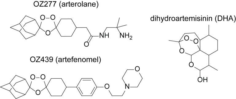
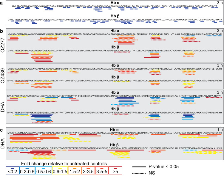
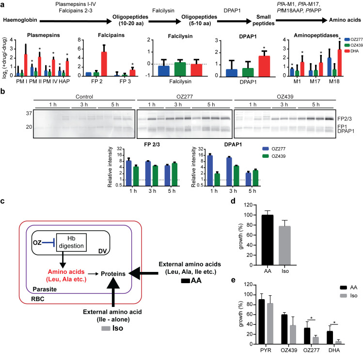
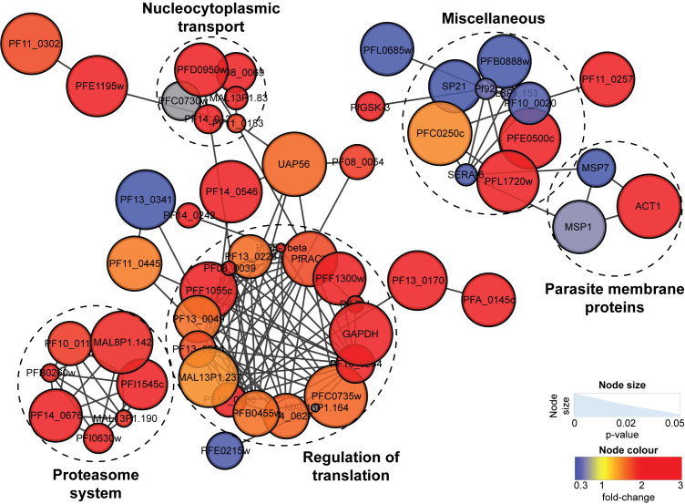
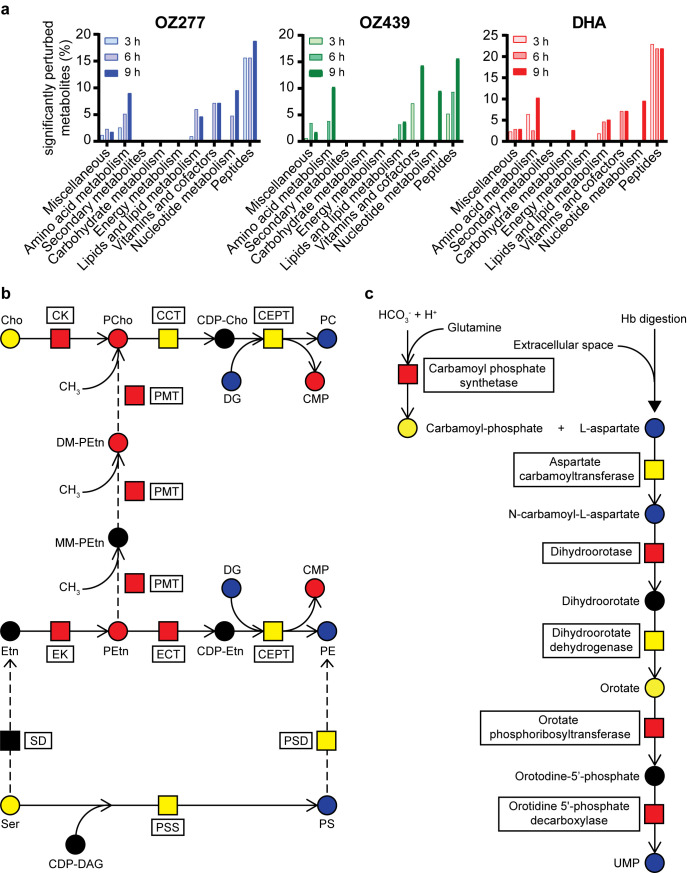
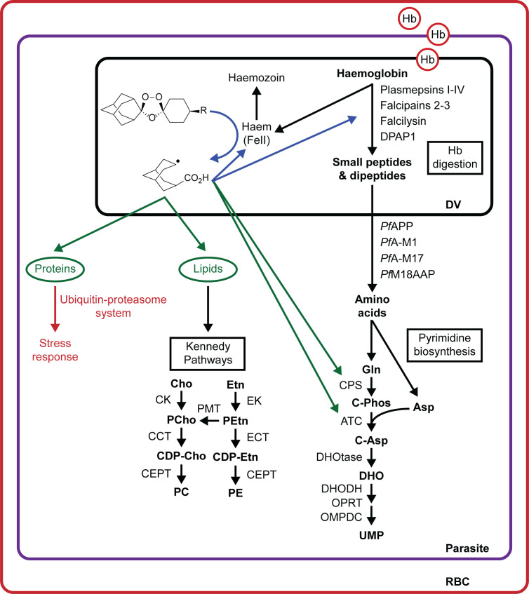
Ozonide antimalarials, OZ277 (arterolane) and OZ439 (artefenomel), are synthetic peroxide-based antimalarials with potent activity against the deadliest malaria parasite, Plasmodium falciparum. Here we used a "multi-omics" workflow, in combination with activity-based protein profiling (ABPP), to demonstrate that peroxide antimalarials initially target the haemoglobin (Hb) digestion pathway to kill malaria parasites. Time-dependent metabolomic profiling of ozonide-treated P. falciparum infected red blood cells revealed a rapid depletion of short Hb-derived peptides followed by subsequent alterations in lipid and nucleotide metabolism, while untargeted peptidomics showed accumulation of longer Hb-derived peptides. Quantitative proteomics and ABPP assays demonstrated that Hb-digesting proteases were increased in abundance and activity following treatment, respectively. Ozonide-induced depletion of short Hb-derived peptides was less extensive in a drug-treated K13-mutant artemisinin resistant parasite line (Cam3.IIR539T) than in the drug-treated isogenic sensitive strain (Cam3.IIrev), further confirming the association between ozonide activity and Hb catabolism. To demonstrate that compromised Hb catabolism may be a primary mechanism involved in ozonide antimalarial activity, we showed that parasites forced to rely solely on Hb digestion for amino acids became hypersensitive to short ozonide exposures. Quantitative proteomics analysis also revealed parasite proteins involved in translation and the ubiquitin-proteasome system were enriched following drug treatment, suggestive of the parasite engaging a stress response to mitigate ozonide-induced damage. Taken together, these data point to a mechanism of action involving initial impairment of Hb catabolism, and indicate that the parasite regulates protein turnover to manage ozonide-induced damage.


Conflict of interest statement

The authors have declared that no competing interests exist.

Figures

Fig 1
Fig 1. Chemical structures of selected peroxide antimalarials.
The fully synthetic ozonide antimalarials, OZ277 (arterolane) and OZ439 (artefenomel), and the clinically used semisynthetic artemisinin derivative, dihydroartemisinin (DHA).
Fig 2
Fig 2. Peroxide-induced perturbations to peptide metabolism.
a, Heatmap showing the average fold change for all identified peptides at each time point after treatment of trophozoite-infected cultures (P. falciparum 3D7 strain) with OZ277, OZ439 and DHA. Values represent the average of at least three biological replicates, expressed relative to the average untreated control value (at least seven biological replicates) for that respective time point. Putative haemoglobin peptides are shown in red text. b, Representative time profiles showing the progressive depletion in abundance of selected putative haemoglobin-derived peptides after peroxide treatment of trophozoite-stage parasites (3D7). Values are the average fold change (± SD) relative to the untreated control of at least three biological replicates. c, Time-dependent decrease in the abundance of the four haemoglobin-derived peptides consistently depleted following peroxide treatment in ring infected cultures (3D7). Values are the average fold change (± SD) relative to the untreated control of four biological replicates. d, Heatmap of the peptides that were altered in abundance (≥ 1.5-fold relative to the untreated control) following peroxide treatment in the K13-wildtype artemisinin sensitive (Cam3.IIrev) and K13-mutant artemisinin resistant (Cam3.IIR539T) parasite lines. Data shown are the average for three or five biological replicates expressed relative to the average for the untreated control from the same parasite line. Putative haemoglobin peptides are shown in red text.
Fig 3
Fig 3. Sequence coverage and relative abundance of haemoglobin alpha (Hb α) and haemoglobin beta (Hb β) peptides detected after peroxide treatment.
a, Sequence coverage for putative haemoglobin-derived di-, tri-, and tetra-peptides that were differentially abundant (≥ 1.5-fold) following peroxide antimalarial treatment relative to the untreated control (3D7 parasites). The peptide sequences PA, PT, PE, PEE, HLD, SLD, PPVQ, PVNF and HVDD have been confirmed by MS/MS analysis. For all other putative peptide sequences, all potential isomers have been mapped. b, Sequence coverage for all long Hb α and Hb β peptides detected in peptidomics studies. Peptide abundances are the average fold change following 3 hours of drug treatment (OZ277, OZ439 and DHA), expressed relative to the untreated control (DMSO) from at least three biological replicates (3D7 parasites). c, Sequence coverage for Hb α and Hb β peptides detected following 1 hour of DHA treatment in Cam3.IIrev (artemisinin sensitive) parasites. Peptide abundances are the average fold change expressed relative to the untreated control (DMSO) from two biological replicates. In b, c, samples were normalised according to peptide concentration (measured using a bicinchoninic acid assay) during sample preparation. The solid lines represent the amino acid sequences of peptides that significantly changed in abundance after drug treatment relative to the untreated control (P-value < 0.05). Dashed lines represent non-significant (NS) changes. Increased and decreased peptide abundance are represented by red and blue (solid or dashed) lines, respectively.
Fig 4
Fig 4. Peroxide antimalarials act by perturbing haemoglobin digestion.
a, Disruption of protease abundance in the haemoglobin digestion pathway [36]. Values are the average log2 fold change (± SD) relative to the untreated control of at least three biological replicates. Trophozoite infected cultures (P. falciparum 3D7) were incubated with OZ277, OZ439 (both 300 nM) or DHA (100 nM) for 3 h. Falcipain 2 (FP 2) was identified in only two OZ277 treatment experiments, therefore the mean alone is shown. Aminopeptidase P (PfAPP) was not identified in any of the proteomic experiments. DPAP1, dipeptidyl aminopeptidase 1; FP, falcipain; HAP, histo-aspartic protease; PfAPP, aminopeptidase P; PfA-M1, alanyl aminopeptidase; PfA-M17, leucyl aminopeptidase; PfM18APP, aspartyl aminopeptidase; PM, plasmepsin; * P-value < 0.05. b, Parasite cysteine protease activity after peroxide treatment using the activity-based probe (ABP), DCG04. Cysteine protease activity and densitometric analysis of the falcipain (FP) 2/3 and dipeptidyl aminopeptidase 1 (DPAP1) signal after OZ277 and OZ439 treatment in P. falciparum trophozoite stage parasites (3D7) using DCG04 in lysates at pH 5.5 (acidic). Trophozoite infected cultures were incubated with OZ277, OZ439 (both 300 nM) or an equivalent volume of DMSO (control). DCG04 labelling was detected by blotting membranes with streptavidin-AF647 after SDS-PAGE and transfer. The lanes for each time point are independent drug treatments and represent at least three biological replicates per time point that were run on the same gel side-by-side. For the densitometric analysis, the post drug treatment FP 2/3 and DPAP1 signal intensity was normalised to the average signal intensity of the appropriate time point in the untreated control (±SD). c, Schematic showing that infected RBCs treated with DHA or ozonides (OZ) can use exogenous amino acids when cultured in AA medium (Full RPMI medium with all 20 amino acids) in response to disrupted haemoglobin digestion (arrow shown in blue), while parasites in Iso medium (supplemented with isoleucine alone at a final concentration of 147.5 μM) must rely solely on haemoglobin digestion for amino acids. d, Amino acid requirement for cultured P. falciparum 3D7 parasites. Parasite viability measured following 48 h incubation in medium containing all amino acids (AA, black bars) and isoleucine alone medium (Iso, grey bars). e, Parasite sensitivity to peroxides when cultured in AA (black bars) medium compared to Iso (grey bars) medium. Trophozoite infected cultures (3D7) were incubated with pyrimethamine (PYR), OZ277, OZ439 (all 300 nM), DHA (100 nM) or an equivalent volume of DMSO (control) for 3 h. Data represents the mean ± SD of at least three biological replicates. Growth values for each treatment is expressed relative to the appropriate untreated medium (Iso or AA) control, which was set to 100%. * P-value < 0.05.
Fig 5
Fig 5. OZ277-induced disruption to the P. falciparum (3D7 strain) proteome.
Network analysis of trophozoite stage parasite proteins perturbed following treatment with OZ277 (300 nM for 3 h). The network analysis was built using the STRINGdb interaction network analysis output (connectivity was based on experimental, database and co-expression evidence with a minimum interaction score of 0.7) in Cytoscape 3.6 with the ClusterONE algorithm. Node size represents P-value and node colour represents fold-change from at least three independent replicates.
Fig 6
Fig 6. Peroxide-induced disruption of secondary biochemical pathways.
a, Metabolic perturbations in trophozoite-stage parasite cultures (P. falciparum 3D7 strain). Pathway enrichment analysis showing the percentage of significantly perturbed metabolites (Welch’s t test; P-value < 0.05 and fold change > 1.5) as a function of metabolite class for extended (3, 6 and 9 h) exposure with OZ277, OZ439 (both 300 nM) and DHA (100 nM). b, Peroxide-induced disruption of the phosphatidylcholine (PC) and phosphatidylethanolamine (PE) lipid biosynthesis pathways within P. falciparum parasites (3D7). The dashed arrows represent an alternative route for the synthesis of PC from ethanolamine (Etn) in P. falciparum. CCT, choline-phosphate cytidyltransferase; CDP-, cytidine-diphospho-; CEPT, choline/ethanolamine phosphotransferase; Cho, choline; CK, choline kinase; CMP, cytidine monophosphate; DAG/DG, diglyceride; DM-, dimethyl-; ECT, ethanolamine-phosphate cytidyltransferase; EK, ethanolamine kinase; Etn, ethanolamine; MM-, monomethyl; PC, phosphatidylcholine; PCho, choline phosphate; PE, phosphatidylethanolamine; PEtn, ethanolamine phosphate; PG, phosphatidylglycerol; PMT, phosphoethanolamine N-methyltransferase; PS, phosphatidylserine; PSD, phosphatidylserine decarboxylase; PSS, phosphatidylserine synthase; SD, serine decarboxylase. c, Peroxide-induced perturbation of pyrimidine biosynthesis in trophozoite-stage parasite cultures (3D7). Hb, haemoglobin; UMP, uridine monophosphate. In b, c, metabolites (circles) and proteins (squares) in red and blue were increased and decreased in abundance after drug treatment, respectively. Yellow and black represent no change and not detected, respectively.
Fig 7
Fig 7. Proposed model for ozonide antimalarial activity in P. falciparum infected red blood cells.
Haemoglobin-derived haem activates peroxide antimalarials within the parasite digestive vacuole. The resulting drug-derived radicals initially damage components proximal to the activation site, including proteases involved in haemoglobin digestion (arrow shown in blue). This leads to disruption of the haemoglobin degradation pathway. To correct for peroxide-induced damage, parasites may respond by increasing the levels of proteases involved in haemoglobin catabolism. Peroxide radicals induce further oxidative insult and cause widespread alkylation of parasite components as the duration of drug exposure is increased (arrows shown in green). This may include damage to lipids, inducing upregulation of the Kennedy Pathways, and proteins involved in other vital parasite functions, such as pyrimidine biosynthesis. To mitigate peroxide-induced cellular damage, the parasite engages a stress response involving translational regulation and the ubiquitin-proteasome systems. Asp, aspartic acid; ATC, aspartate carbamoyltransferase; C-Asp, carbamoyl-aspartate, C-Phos, carbamoyl-phosphate; CCT, choline-phosphate cytidyltransferase; CDP-, cytidine-diphospho-; CEPT, choline/ethanolamine phosphotransferase; Cho, choline; CK, choline kinase; CMP, cytidine monophosphate; CPS, carbamoyl phosphate synthetase; DHO, dihydroorotate; DHODH, dihydroorotate dehydrogenase; DHOtase, dihydroorotase; DV, digestive vacuole; ECT, ethanolamine-phosphate cytidyltransferase; EK, ethanolamine kinase; Etn, ethanolamine; Gln, glutamine; Hb, haemoglobin; OMPDC, orotodine 5’-phosphate decarboxylase; OPRT, orotate phosphoribosyltransferase; PC, phosphatidylcholine; PE, phosphatidylethanolamine; PCho, choline phosphate; PEtn, ethanolamine phosphate; PMT, phosphoethanolamine N-methyltransferase; RBC, red blood cell; UMP, uridine monophosphate.

Similar articles

Cited by

References

    1. WHO. World Malaria Report 2017. Geneva: World Health Organisation, 2017.
    1. Enserink M. Malaria's drug miracle in danger. Science. 2010; 328: 844–6. 10.1126/science.328.5980.844 . - DOI - PubMed
    1. Meshnick SR, Thomas A, Ranz A, Xu CM, Pan HZ. Artemisinin (qinghaosu): the role of intracellular hemin in its mechanism of antimalarial action. Mol Biochem Parasitol. 1991; 49: 181–9. 10.1016/0166-6851(91)90062-b . - DOI - PubMed
    1. Meshnick SR, Yang YZ, Lima V, Kuypers F, Kamchonwongpaisan S, Yuthavong Y. Iron-dependent free radical generation from the antimalarial agent artemisinin (qinghaosu). Antimicrob Agents Chemother. 1993; 37: 1108–14. 10.1128/aac.37.5.1108 . - DOI - PMC - PubMed
    1. Posner GH, Oh CH. Regiospecifically oxygen-18 labeled 1, 2, 4-trioxane: a simple chemical model system to probe the mechanism (s) for the antimalarial activity of artemisinin (qinghaosu). J Am Chem Soc. 1992; 114: 8328–9. 10.1021/ja00047a076 - DOI

Publication types

MeSH terms