Case Reports
. 2020 Aug;61(8):e90-e94.
doi: 10.1111/epi.16612. Epub 2020 Jul 23.

Orbitofrontal involvement in a neuroCOVID-19 patient

Affiliations
Case Reports

Orbitofrontal involvement in a neuroCOVID-19 patient

Loïc Le Guennec et al. Epilepsia. 2020 Aug.

Abstract

Neurological manifestations of coronavirus disease 19 (COVID-19) such as encephalitis and seizures have been reported increasingly, but our understanding of COVID-19-related brain injury is still limited. Herein we describe prefrontal involvement in a patient with COVID-19 who presented prior anosmia, raising the question of a potential trans-olfactory bulb brain invasion.


Conflict of interest statement

The authors have no conflicts of interest to declare in relationship with this manuscript. We confirm that we have read the Journal's position on issues involved in ethical publication and affirm that this report is consistent with those guidelines.

Figures
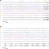
FIGURE 1
FIGURE 1
Electroencephalography(EEG) findings. Samples of a scalp EEG recording, in a bipolar montage, showing (A) repetitive 1 Hz rhythmic bursts predominating on the right frontal lobe, suggestive of a nonconvulsive status epilepticus and (B) 2 days later, lateralized periodic discharges (LPDs) on the right frontal lobe
FIGURE 2
FIGURE 2
Brain magnetic resonance imaging (MRI). Upper panel: Brain MRI on admission: A and B, Axial diffusion‐weighted imaging (DWI) showing right orbitofrontal cortex and caudate nucleus hyperintensity (tip of the red arrow). C, Sagittal fluid‐attenuated inversion recovery (T2/FLAIR) showing right orbitofrontal cortico‐subcortical hyperintensity toward the caudate nucleus (tip of the red arrow). D, Coronal T2/FLAIR showing an orbitofrontal hyperintensity (tip of the red arrow). Lower panel: Brain MRI on day 15: E and F, Axial DWI showing decrease of the hyperintensity within the right orbitofrontal cortex but persistence on the right caudate nucleus (tip of the red arrow). G, Sagittal and H, coronal T2/FLAIR showing decrease of the hyperintensity within the right orbitofrontal cortex

References

    1. Mao L, Jin H, Wang M, Hu YU, Chen S, He Q, et al. Neurologic manifestations of hospitalized patients with coronavirus disease 2019 in Wuhan, China. JAMA Neurol. 2020;77(6):683. - PMC - PubMed
    1. Helms J, Kremer S, Merdji H, Clere‐Jehl R, Schenck M, Kummerlen C, et al. Neurologic features in severe SARS‐CoV‐2 infection. N Engl J Med. 2020;382(23):2268–70. - PMC - PubMed
    1. Moriguchi T, Harii N, Goto J, Harada D, Sugawara H, Takamino J, et al. A first case of meningitis/encephalitis associated with SARS‐Coronavirus‐2. Int J Infect Dis. 2020;94:55–8. - PMC - PubMed
    1. Poyiadji N, Shahin G, Noujaim D, Stone M, Patel S, Griffith B. COVID‐19‐associated acute hemorrhagic necrotizing encephalopathy: CT and MRI features. Radiology. 2020;31:2020201187. - PMC - PubMed
    1. Filatov A, Sharma P, Hindi F, Espinosa PS. Neurological complications of coronavirus disease (COVID‐19): encephalopathy. Cureus. 2020;12:e7352. - PMC - PubMed

Publication types