. 2020 Jul 23;182(2):345-356.e16.
doi: 10.1016/j.cell.2020.06.005. Epub 2020 Jun 25.

Recognition of Semaphorin Proteins by P. sordellii Lethal Toxin Reveals Principles of Receptor Specificity in Clostridial Toxins

Affiliations

Recognition of Semaphorin Proteins by P. sordellii Lethal Toxin Reveals Principles of Receptor Specificity in Clostridial Toxins

Hunsang Lee et al. Cell. .

Abstract

Pathogenic clostridial species secrete potent toxins that induce severe host tissue damage. Paeniclostridium sordellii lethal toxin (TcsL) causes an almost invariably lethal toxic shock syndrome associated with gynecological infections. TcsL is 87% similar to C. difficile TcdB, which enters host cells via Frizzled receptors in colon epithelium. However, P. sordellii infections target vascular endothelium, suggesting that TcsL exploits another receptor. Here, using CRISPR/Cas9 screening, we establish semaphorins SEMA6A and SEMA6B as TcsL receptors. We demonstrate that recombinant SEMA6A can protect mice from TcsL-induced edema. A 3.3 Å cryo-EM structure shows that TcsL binds SEMA6A with the same region that in TcdB binds structurally unrelated Frizzled. Remarkably, 15 mutations in this evolutionarily divergent surface are sufficient to switch binding specificity of TcsL to that of TcdB. Our findings establish semaphorins as physiologically relevant receptors for TcsL and reveal the molecular basis for the difference in tissue targeting and disease pathogenesis between highly related toxins.

Keywords: CRISPR/Cas9 screening; Clostridial toxins; P. sordellii; SEMA6A; SEMA6B; TcsL; bacterial exotoxins; cryo-EM; semaphorin.


Conflict of interest statement

Declaration of Interests The authors declare no competing interests.

Figures

None
Graphical abstract
Figure 1
Figure 1
SEMA6A and SEMA6B are host cell receptors for P. sordellii lethal toxin TcsL (A) Genome-wide CRISPR/Cas9 screen in Hap1 cells identifies factors regulating sensitivity to 0.1 nM TcsL. Hap1 cells were infected with a genome-wide TKOv3 gRNA library, treated with recombinant TcsL, and gRNAs from surviving cells were sequenced. (B) Genome-wide CRISPR/Cas9 screen with 1 nM TcsL. (C) Phylogenetic tree of SEMA6 family proteins. (D) Hap1 cells were infected with Cas9 and gRNA targeting indicated genes and tested for sensitivity to TcsL. Data (n = 3) are represented as mean ± standard deviation. Shown at the bottom, expression of SEMA6A and SEMA6B in single and double knockout cell lines was assessed by western blotting. (E) Hap1 SEMA6AKO cells were infected with lentiviruses expressing 3xFLAG-tagged SEMA6 family proteins and tested for TcsL sensitivity. Data (n = 3) are represented as mean ± standard deviation. Shown at the bottom, expression of SEMA6 proteins in infected cell lines was validated with western blotting. See also Figure S1 and Table S1.
Figure S1
Figure S1
Validation of SEMA6A and SEMA6B as host factors required for TcsL intoxication, related to Figure 1 and Figure 2 (A) Expression of SEMA6 family genes, the cognate SEMA6A/6B ligands Plexin A2 and Plexin A4, and known clostridial toxin receptors and host cell factors in Hap1 and HeLa cells based on Human Protein Atlas (proteinatlas.org). (B) Left, Sensitivity of SEMA6A and UGP2 knockout cells to TcsL. SEMA6A and UGP2 knockout cells were generated with CRISPR/Cas9. SEMA6A-3xFLAG was ectopically expressed in SEMA6A knockout cells by lentiviral infection. Data (n = 3) are represented as mean ± standard deviation. Right, SEMA6A expression in wild-type Hap1 cells, SEMA6AKO cells, and in SEMA6AKO cells ectopically expressing SEMA6A-3xFLAG. (C) Hap1 and HeLa cells were treated with increasing concentrations of TcsL and cell viability measured 24 h later. Data (n = 3) are represented as mean ± standard deviation. Inset, expression of SEMA6A in Vero, Hap1, and HeLa cells was assessed by western blot. (D) SEMA6A ectodomain protects Vero cells from TcsL toxicity only when added simultaneously with the toxin. SEMA6A and TcsL were added to Vero cells simultaneously or after 1-min or 1 h pre-incubation. Alternatively, TcsL was added for 1 h prior to treatment with SEMA6A. Data (n = 2) are represented as mean ± standard deviation
Figure 2
Figure 2
SEMA6A and SEMA6B ectodomains protect cells from TcsL intoxication (A) Vero cells stably expressing Nanoluciferase viability reporter were treated with increasing amounts of TcsL in the presence of recombinant SEMA6A ectodomain. Data (n = 2) are represented as mean ± standard deviation. (B) Vero cells stably expressing Nanoluciferase viability reporter were treated with 50 pM TcsL with increasing amounts of recombinant SEMA6 family ectodomains for 24 h. Data (n = 2) are represented as mean ± standard deviation. (C) Microscopy images of Vero cells treated with TcsL (50 pM) and SEMA6 family ectodomains (1 uM) for 24 h. See also Figure S1.
Figure 3
Figure 3
SEMA6A ectodomain protects lung endothelial cells and mouse lungs from TcsL-induced toxicity (A) SEMA6A and SEMA6B protein expression in human lung endothelial cells. (B) HULECs are extremely sensitive to TcsL. HULEC-5a cells were treated with increased amounts of indicated Clostridial toxins. (C) Recombinant mouse Sema6a ectodomain fused to Fc domain protects HULEC-5a cells from TcsL-induced cell rounding. Cells were treated with 5 pM TcsL and increasing amounts of Sema6a and Sema6c ectodomains. (D) Microscopy images of HULEC-5a cells treated with TcsL and recombinant Sema6a and Sema6c ectodomains. (E) Immunohistochemistry images of Sema6a and Sema6b expression in mouse lung tissue sections. (F) Mice were intraperitoneally injected with 15 ng TcsL and 1,000-fold molar excess of Sema6a ectodomain, Sema6c ectodomain or BSA. Thoracic fluid was collected and measured from symptomatic mice 4 h after injection. (G) Lung tissue sections of mice treated with indicated conditions. Arrows indicate lung edema induced by TcsL.
Figure 4
Figure 4
Cryo-EM structure of the TcsL1285–1804-SEMA6A complex (A) On the left, domain structure of TcsL and SEMA6A. Constructs used in cryo-EM analysis are indicated below. Darker blue color indicates TcsL region resolved to medium and high resolution (< 7 Å) in cryo-EM. Shown on the right is a schematic indicating the location of the TcsL fragment used for cryo-EM, based on full-length TcdB structure (PDB: 6OQ5). Abbreviations are as follows: GTD, glucosyltransferase domain; APD, autoprocessing domain; delivery, delivery domain; CROP, combined repetitive oligopeptides; SEMA, semaphorin domain, PSI, plexin-semaphorin-integrin domain; TMD, transmembrane domain. (B) Composite cryo-EM map of the Tcsl-SEMA6A complex. SEMA6A monomers are colored pink and yellow, TcsL domain resolved to medium and high resolution (< 7 Å) is colored dark blue. Low-pass filtered density (10 Å) of the TcsL protein is shown in light blue. Insets I and II show contact residues between SEMA6A (pink) and TcsL (blue). (C) Atomic model built into the SEMA6A-TcsL map. Cryo-EM density of SEMA6A-TcsL is shown as gray mesh with the model built shown as sticks (pink for SEMA6A and blue for TcsL). (D) Comparison of Plexin A2-SEMA6A (PDB: 3OKY) (Janssen et al., 2010) and TcsL-SEMA6A binding interactions. M109 of SEMA6A interacts with hydrophobic residues of Plexin A2, including L407 and V398 (left). TcsL buries M109 of SEMA6A in a binding pocket containing several hydrophobic residues (middle). An overlay of TcsL and Plexin A2 binding surfaces (shown in blue and purple, respectively) reveals a subset of SEMA6A residues participating in binding to both protein ligands (red). See also Figure S2, Figure S3, Figure S4, Figure S5.
Figure S3
Figure S3
Biochemical and cryo-EM analysis of the SEMA6A/TcsL complex, related to Figure 4 and Figure 5 (A) Representative binding curves for the TcsL1285-1804/SEMA6A interaction. In this analysis, His-tagged TcsL1285-1804 was immobilized on the Ni-NTA biosensor. The average apparent binding affinity from four independent experiments is 2.4 ± 0.8 nM. The data (blue) were fitted using a 1:1 binding model (red). (B) An example of cryo-EM micrograph. Scale bar is 50 nm. (C) Selected 2D class averages of the TcsL/SEMA6A complex. (D) Gold standard Fourier shell correlation (GSFSC) curve of the final 3D non-uniform refinement of the TcsL/SEMA6A complex in cryoSPARC v2. (E) Viewing direction distribution of the TcsL/SEMA6A data. (F) Selected 2D class averages of SEMA6A dimer. (G) GSFSC curve of the final 3D non-uniform refinement of the SEMA6A dimer in cryoSPARC v2. (H) Viewing direction distribution of the SEMA6A dimer data.
Figure S2
Figure S2
Cryo-EM data processing workflow in cryoSPARC v2, related to Figure 4 and Figure 5
Figure S4
Figure S4
Local resolution (Å) plotted on the surface of cryo-EM map of the SEMA6A/TcsL complex and SEMA6A dimer, related to Figure 4 and Figure 5 (A) Local resolution of the SEMA6A/TcsL ranges from 2.8 Å at the core of the SEMA6A to > 30 Å at the flexible terminus of TcsL. (B) Local resolution plotted on the surface of TcsL (left) and SEMA6A (right) binding interfaces. (C) Local resolution plotted on the surface of the SEMA6A cryo-EM map.
Figure S5
Figure S5
Comparison of published structures of clostridial toxins and SEMA6A, related to Figure 4 and Figure 5 (A) Left, Cryo-EM structure of TcsL toxin fragment (residues in construct: 1283-1804, residues resolved and modeled: 1400-1637); middle, X-ray structure of TcdB toxin fragment (residues in the model 1283-1804; PDB ID: 6C0B; Chen et al., 2018); right, X-ray structure of TcdA toxin fragment (residues in the model 1-1832; PDB ID: 4R04; Chumbler et al., 2016). Binding interfaces of TcsL and TcdB to their respective receptors are highlighted in light blue. “H” and “S” letters denote the positions of selected α helices and β strands conserved in all three toxin structures. The glucosyltransferase domain is highlighted in pink, the autoprotease domain is highlighted in green and the delivery domains are highlighted in gray. (B) Comparison of SEMA6A in different structures. All atom RMSD values were calculated using Pymol and are plotted on the surface of SEMA6A dimers (left) and monomers (right).
Figure S6
Figure S6
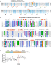
Conservation of SEMA6A and TcsL interface residues in semaphoring and large clostridial toxin families, related to Figure 6 A, Sequence alignment of SEMA6 family proteins. Residues conserved in all four SEMA6 family proteins are denoted in light blue. SEMA6A residues forming contacts with TcsL are indicated as red circles, and those interacting with Plexin A2 are indicated as blue circles. The two TcsL-interacting residues that differ between SEMA6A/SEMA6B and SEMA6C/SEMA6D are colored orange. Secondary structure elements with numbered beta-propeller blades are shown below the alignment. (B) The receptor-binding surface of TcsL and TcdB is highly divergent between all clostridial toxins. Partial sequence alignment of six known large clostridial toxins. Secondary structure elements and the consensus sequence (at least 4/6 identical residues) are shown above the alignment. Amino acids are colored based on their biophysical properties (ClustalX coloring) if at least 4/6 residues are similar in each column. Red boxes indicate interface residues in TcsL/SEMA6A or TcdB/Fzd2 complexes. The gray box highlights the evolutionarily divergent beta sheet in the receptor-binding interface. (C) Alignment entropy was calculated as a 20-aa moving window along the alignment of six known clostridial toxins. Red bars indicate the location of TcsL/SEMA6A interface residues.
Figure 5
Figure 5
Clostridial toxins use the same region to bind their cognate receptors (A) Comparison of the TcsL-SEMA6A and TcdB-FZD2 (PDB:6C0B) (Chen et al., 2018) binding interface. Residues mutated in TcsL4mut are indicated in red. (B) TcsL buries M109 of SEMA6A in a hydrophobic binding pocket (left), whereas TcdB utilizes a similar hydrophobic binding pocket to interact with the palmitoleic acid moiety of FZD2 (right). (C) Experimental validation of the TcsL-SEMA6A interaction interface. The cytotoxicity of wild-type TcsL and TcsL4mut variant with four mutated interaction interface residues (C1433D-I1434K-A1486S-Y1596R; shown in A) was assessed in Vero cells. (D) Validation of SEMA6A M109 as a critical interacting residue with TcsL. SEMA6A/SEMA6B double knockout cells were infected with 3xFLAG-tagged wild-type SEMA6A or M109D mutant and assayed for sensitivity to TcsL. Protein expression levels were confirmed by western blotting (right). See also Figure S2, Figure S3, Figure S4, Figure S5.
Figure 6
Figure 6
TcsL and TcdB bind different host receptors through the same interface region (A) Sequence alignment entropy in large clostridial toxin family shown as a rainbow spectrum on the TcdB full-length cryo-EM structure (PDB ID: 6OQ5) (Chen et al., 2019). Entropy was calculated as a 10-aa moving window. The receptor-binding surface is indicated. (B) SEC profiles and SDS-PAGE analysis of FZD7-TcdB1285–1804 (top), SEMA6A-TcsL1285–1804 (middle), and FZD7-TcsL1285–1804 (bottom). SEC fractions used for SDS-PAGE analysis are highlighted with an asterisk. (C) Sequence alignment between TcsL and TcsB. TcsL residues interacting with SEMA6A are highlighted in pink and TcdB residues contacting FZD2 are highlighted in orange. Black dots denote the 15 mutations introduced in TcsL (FBD)1285–1804 variant that resulted in shifting the TcsL binding specificity from SEMA6A to FZD7. (D) SEC profiles and SDS-PAGE analysis of FZD7-TcsL (FBD)1285–1804 (TcsL variant with a TcdB-like binding interface) (top) and SEMA6A-TcsL (FBD)1285–1804 (bottom). SEC fractions used for SDS-PAGE analysis are highlighted with an asterisk. See also Figure S6.

Comment in

Similar articles

Cited by

References

    1. Adams P.D., Afonine P.V., Bunkóczi G., Chen V.B., Davis I.W., Echols N., Headd J.J., Hung L.-W., Kapral G.J., Grosse-Kunstleve R.W. PHENIX: a comprehensive Python-based system for macromolecular structure solution. Acta Crystallogr. D Biol. Crystallogr. 2010;66:213–221. - PMC - PubMed
    1. Aktories K., Barbieri J.T. Bacterial cytotoxins: targeting eukaryotic switches. Nat. Rev. Microbiol. 2005;3:397–410. - PubMed
    1. Aktories K., Just I. Monoglucosylation of low-molecular-mass GTP-binding Rho proteins by clostridial cytotoxins. Trends Cell Biol. 1995;5:441–443. - PubMed
    1. Aldape M.J., Bayer C.R., Bryant A.E., Stevens D.L. A novel murine model of Clostridium sordellii myonecrosis: Insights into the pathogenesis of disease. Anaerobe. 2016;38:103–110. - PMC - PubMed
    1. Carter G.P., Awad M.M., Hao Y., Thelen T., Bergin I.L., Howarth P.M., Seemann T., Rood J.I., Aronoff D.M., Lyras D. TcsL is an essential virulence factor in Clostridium sordellii ATCC 9714. Infect. Immun. 2011;79:1025–1032. - PMC - PubMed

Publication types

MeSH terms