Review
. 2020:140:317-340.
doi: 10.1016/bs.ctdb.2019.10.006. Epub 2019 Nov 19.

Maternal factors regulating preimplantation development in mice

Affiliations
Review

Maternal factors regulating preimplantation development in mice

Di Wu et al. Curr Top Dev Biol. 2020.

Abstract

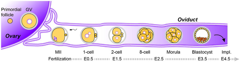
Mammalian embryogenesis depends on maternal factors accumulated in eggs prior to fertilization and on placental transfers later in gestation. In this review, we focus on initial events when the organism has insufficient newly synthesized embryonic factors to sustain development. These maternal factors regulate preimplantation embryogenesis both uniquely in pronuclear formation, genome reprogramming and cell fate determination and more universally in regulating cell division, transcription and RNA metabolism. Depletion, disruption or inappropriate persistence of maternal factors can result in developmental defects in early embryos. To better understand the origins of these maternal effects, we include oocyte maturation processes that are responsible for their production. We focus on recent publications and reference comprehensive reviews that include earlier scientific literature of early mouse development.

Keywords: Maternal RNA degradation; Preimplantation embryonic development; Zygotic genome activation.


Figures

Fig. 1
Fig. 1
An outline of mouse oogenesis and preimplantation development. Oocytes grow and undergo meiotic maturation within the ovary prior to ovulation into the oviduct where they are fertilized by capacitated sperm to form a zygote (embryonic day 1, E1). The 1-cell zygote divides into two totipotent blastomeres and continues to cleave (~E1.5). At the 8-cell stage (~E2.5), the embryo compacts to form the morula. After 2–3 additional rounds of cell division (~E3.5), a blastocoel forms, transforming the embryo into a blastocyst that implants on the wall of the uterus ~E4.5. Impl., Implantation.
Fig. 2
Fig. 2
Maternal factors function in chromatin remodeling post-fertilization. (A) Paternal pronucleus formation by maternal NPM2 that mediates protamine removal and H3.3 that mediates recondensation. Maternal chromatin is occupied by H3.3 and H3.1/3.2. (B) Paternal DNA demethylation is mediated by maternal TET proteins while maternal DNA methylation persists due to the masking by DPPA3. (C) DNA methylation differs in paternal and maternal alleles during preimplantation development. Paternal DNA is actively and rapidly demethylated at the 1-cell stage whereas maternal DNA methylation decreases passively through cell division. Both paternal and maternal DNA then undergo remethylation by DNMT3A/3B beginning in blastocysts. Imprinted genes remain unaffected by DNMT1 during preimplantation. (D) Establishment of topologically associating domains (TADs) during preimplantation. While there are no clearly defined TADs at the 1-cell stage, they are gradually established during preimplantation development.
Fig. 3
Fig. 3
Maternal factors affect zygotic gene activation (ZGA). (A) Minor and major waves of ZGA in the 1- and 2-cell embryos. (B) Maternal factors can directly regulate ZGA by affecting transcription (red) or chromatin structure (blue), or indirectly regulate ZGA by affecting other cellular activities including RNA metabolism, DNA repair and cytoskeleton (black).
Fig. 4
Fig. 4
Gene network in cell fate specification and differentiation in morula-to-blastocyst transition. (A) SOX2 and CDX2 determine inner-outer polarity at the morula stage. HIPPO pathway modulates the SOX2 and CDX2 balance through interacting with Notch and Rho pathways. The inner-outer polarity is maintained by a SOX2-NANOG-OCT4 network in the inner cell mass (ICM), which is inhibited by CDX2 in the trophectoderm (TE). (B) The established inner-outer polarity persists and the ICM differentiates by priming NANOG and GATA6 to form the epiblast and primitive endoderm which are regulated by zygotic FGF signaling.
Fig. 5
Fig. 5
Maternal transcriptome shaping and maternal RNA clearance. (A) Maternal transcriptome shaping happens during oocyte maturation from GV to MII stages. The protein coding transcripts can be either polyadenylated for translation (red), or deadenylated. The deadenylated RNAs can be degraded (blue) or protected in RNA-containing granules (magenta). (B) Maternal RNA clearance happens post-fertilization to ensure zygotic control of subsequent developmental programs. The egg-deposited RNAs can be either polyadenylated for translation, or deadenylated for degradation.

References

    1. Aoki F, Worrad DM, & Schultz RM (1997). Regulation of transcriptional activity during the first and second cell cycles in the preimplantation mouse embryo. Developmental Biology, 181, 296–307. - PubMed
    1. Avilion AA, Nicolis SK, Pevny LH, Perez L, Vivian N, & Lovell-Badge R (2003). Multipotent cell lineages in early mouse development depend on SOX2 function. Genes & Development, 17, 126–140. - PMC - PubMed
    1. Balhorn R (2007). The protamine family of sperm nuclear proteins. Genome Biology, 8, 227. - PMC - PubMed
    1. Baran V, Solc P, Kovarikova V, Rehak P, & Sutovsky P (2013). Polo-like kinase 1 is essential for the first mitotic division in the mouse embryo. Molecular Reproduction and Development, 80, 522–534. - PubMed
    1. Bebbere D, Masala L, Albertini DF, & Ledda S (2016). The subcortical maternal complex: Multiple functions for one biological structure? Journal of Assisted Reproduction and Genetics, 33, 1431–1438. - PMC - PubMed

Publication types

LinkOut - more resources