

Surface-guided tomotherapy improves positioning and reduces treatment time: A retrospective analysis of 16 835 treatment fractions
- PMID: 32592288
- PMCID: PMC7484821
- DOI: 10.1002/acm2.12936
Surface-guided tomotherapy improves positioning and reduces treatment time: A retrospective analysis of 16 835 treatment fractions
Abstract
Purpose: In this study, we have quantified the setup deviation and time gain when using fast surface scanning for daily setup/positioning with weekly megavoltage computed tomography (MVCT) and compared it to daily MVCT.
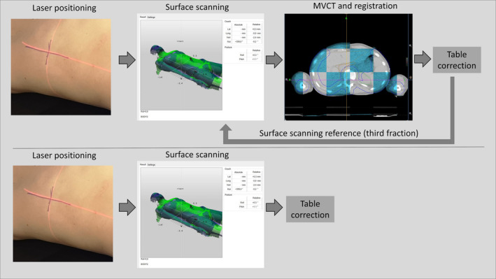
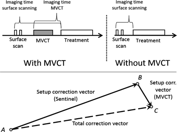
Methods: A total of 16 835 treatment fractions were analyzed, treated, and positioned using our TomoTherapy HD (Accuray Inc., Madison, USA) installed with a Sentinel optical surface scanning system (C-RAD Positioning AB, Uppsala, Sweden). Patients were positioned using in-room lasers, surface scanning and MVCT for the first three fractions. For the remaining fractions, in-room laser was used for setup followed by daily surface scanning with MVCT once weekly. The three-dimensional (3D) setup correction for surface scanning was evaluated from the registration between MVCT and the planning CT. The setup correction vector for the in-room lasers was assessed from the surface scanning and the MVCT to planning CT registration. The imaging time was evaluated as the time from imaging start to beam-on.
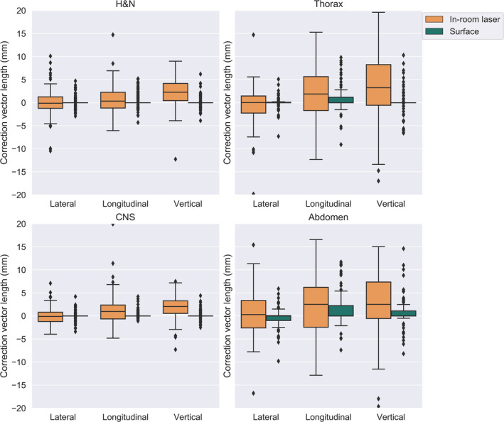
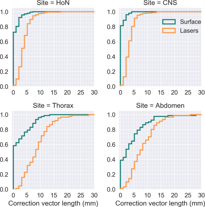
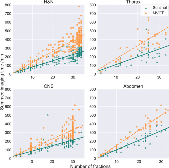
Results: We analyzed 894 TomoTherapy treatment plans from 2012 to 2018. Of all the treatment fractions performed with surface scanning, 90 % of the residual errors were within 2.3 mm for CNS (N = 284), 2.9 mm for H&N (N = 254), 8.7 mm for thorax (N = 144) and 10.9 for abdomen (N = 134) patients. The difference in residual error between surface scanning and positioning with in-room lasers was significant (P < 0.005) for all sites. The imaging time was assessed as total imaging time per treatment plan, modality, and treatment site and found that surface scanning significantly reduced patient on-couch time compared to MVCT for all treatment sites (P < 0.005).
Conclusions: The results indicate that daily surface scanning with weekly MVCT can be used with the current target margins for H&N, CNS, and thorax, with reduced imaging time.
Keywords: SGRT; helical; radiotherapy; surface scanning; tomotherapy.
© 2020 The Authors. Journal of Applied Clinical Medical Physics published by Wiley Periodicals, Inc. on behalf of American Association of Physicists in Medicine.
Conflict of interest statement
The authors have no relevant conflict of interest.
Figures
References
-
- Guckenberger M, Meyer J, Vordermark D, Baier K, Wilbert J, Flentje M. Magnitude and clinical relevance of translational and rotational patient setup errors: a cone‐beam CT study. Int J Radiat Oncol Biol Phys. 2006;65:934–942. - PubMed
-
- Landberg T, Chavaudra J, Dobbs J, et al Report 50. J Int Comm Radiat Units Meas. 1993;os26:NP.
-
- Mackie TR, Holmes T, Swerdloff S et al Tomotherapy: a new concept for the delivery of dynamic conformal radiotherapy. Med Phys. 1993;20:1709–1719. - PubMed
-
- Accuray I . TOMOTHERAPY® H™ SERIES: TomoH™, TomoHD™ and TomoHDA™ Systems Technical Specifications. In: Accuray I, ed. Madison, WI, US; 2017.
-
- Mackie TR, Kapatoes J, Ruchala K, et al Image guidance for precise conformal radiotherapy. Int J Radiat Oncol Biol Phys. 2003;56:89–105. - PubMed
MeSH terms
LinkOut - more resources
Full Text Sources
