. 2020 Aug;10(8):1668-1684.
doi: 10.1002/2211-5463.12919. Epub 2020 Jul 22.

Biochemical determinants of the IGFBP-3-hyaluronan interaction

Affiliations

Biochemical determinants of the IGFBP-3-hyaluronan interaction

Sadaf Dorandish et al. FEBS Open Bio. 2020 Aug.

Abstract

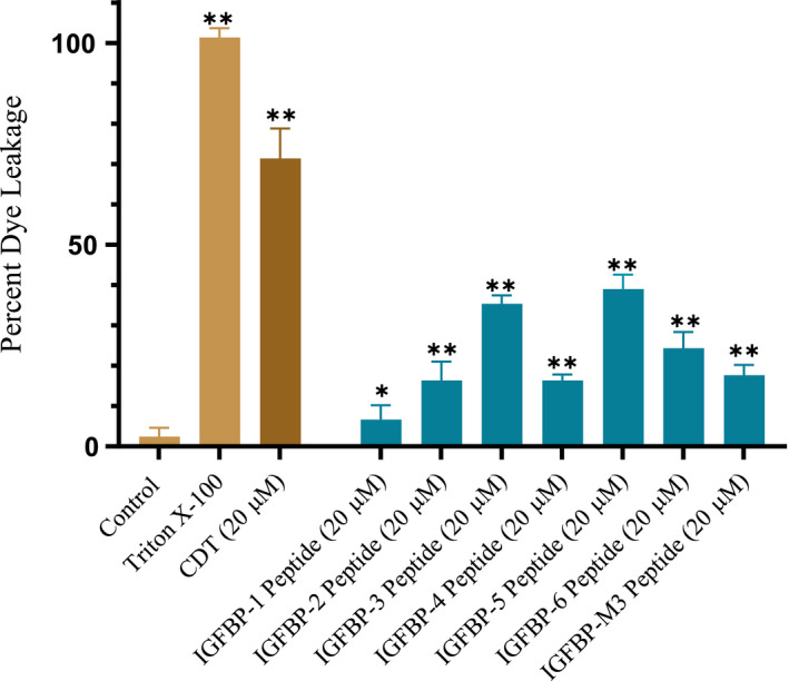
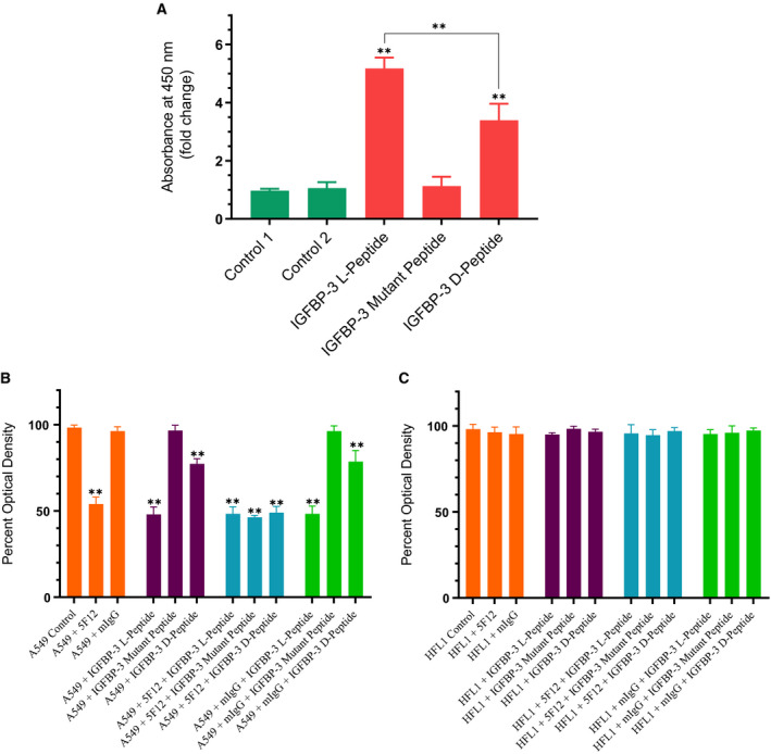
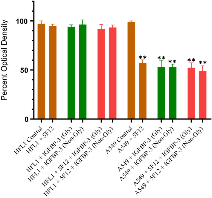
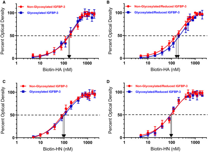
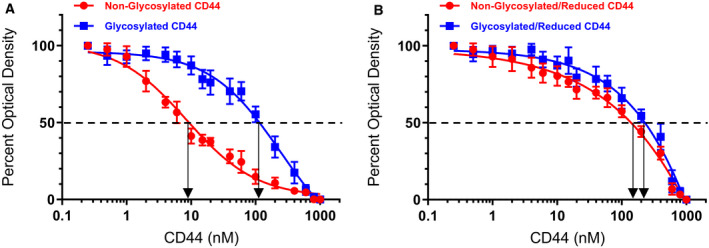
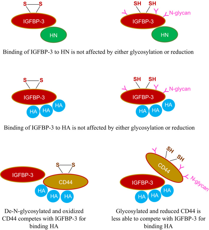
IGFBP-3, the most abundant IGFBP and the main carrier of insulin-like growth factor I (IGF-I) in the circulation, can bind IGF-1 with high affinity, which attenuates IGF/IGF-IR interactions, thereby resulting in antiproliferative effects. The C-terminal domain of insulin-like growth factor-binding protein-3 (IGFBP-3) is known to contain an 18-basic amino acid motif capable of interacting with either humanin (HN) or hyaluronan (HA). We previously showed that the 18-amino acid IGFBP-3 peptide is capable of binding either HA or HN with comparable affinities to the full-length IGFBP-3 protein and that IGFBP-3 can compete with the HA receptor, CD44, for binding HA. Blocking the interaction between HA and CD44 reduced viability of A549 human lung cancer cells. In this study, we set out to better characterize IGFBP-3-HA interactions. We show that both stereochemistry and amino acid identity are important determinants of the interaction between the IGFBP-3 peptide and HA and for the peptide's ability to exert its cytotoxic effects. Binding of IGFBP-3 to either HA or HN was unaffected by glycosylation or reduction of IGFBP-3, suggesting that the basic 18-amino acid residue sequence of IGFBP-3 remains accessible for interaction with either HN or HA upon glycosylation or reduction of the full-length protein. Removing N-linked oligosaccharides from CD44 increased its ability to compete with IGFBP-3 for binding HA, while reduction of CD44 rendered the protein relatively ineffective at blocking IGFBP-3-HA interactions. We conclude that both deglycosylation and disulfide bond formation are important for CD44 to compete with IGFBP-3 for binding HA.

Keywords: CD44; IGFBP-3; humanin; hyaluronan; kinetics; peptide.


Conflict of interest statement

All authors read and approved the final manuscript and declare no conflict of interest with the contents of this article. ‘The content is solely the responsibility of the authors and does not necessarily represent the official views of the National Institutes of Health’.

Figures

Fig. 1
Fig. 1
The IGFBP‐3 peptide has less liposome disruption capability compared to the CDT control. Liposomes were prepared as described in Materials and methods followed by addition of the IGFBP peptides. Negative controls contained 5% DMSO/PBS, which was predetermined not to cause dye leakage, while Triton X‐100 and CDT were used as positive controls. After a 10‐min period, fluorescence values of the samples in 96‐well plates were measured by a spectrofluorometer (filter set to 485 nm excitation and 528 nm emission). Fluorescence measurements indicate dye leakage corresponding to liposome damage. Triton X‐100 detergent (10% v/v in PBS) was used as the positive control for determination of 100% leakage. Percent liposome dye leakage was calculated, and each column represents the mean ± SD of three independent experiments, each run in triplicate. The asterisks (*P < 0.05, **P < 0.01) indicate a statistically significant difference from the control. The absence of asterisks indicates no significance, Mann–Whitney test.
Fig. 2
Fig. 2
WT IGFBP‐3 l‐peptide is more effective than the d‐peptide in both binding HA and in blocking viability of A549 cells that express CD44 as compared to the CD44‐negative cell line, HFL1. (A) IGFBP‐3 peptides (50 nm each) were bound to the ELISA plate wells, and then, 200 nm biotin‐HA was added and processed as described in the Materials and methods section. The data were normalized to the control incubated with BSA (control 1), and fold change relative to the control was calculated. Control 2 is a negative control that included bound WT IGFBP‐3 l‐peptide and added streptavidin–HRP and TMB without addition of biotin‐HA. Each column represents the mean ± SD of three independent experiments, each run in triplicate. The asterisks (**P < 0.01) indicate a statistically significant difference from control 1 and of the IGFBP‐3 l‐peptide compared to the d‐counterpart. The absence of asterisks indicates no significance, Mann–Whitney test. (B, C) IGFBP‐3 peptides were added to cells in the absence or presence of the mIgG (5 μg·mL−1) antibody control or the CD44, 5F12 antibody (5 μg·mL−1), known to block HA‐CD44 interactions. Cell viability was assessed by the MTT assay. Cells were seeded in 96‐well plates at 0.2 × 105 cells per well in 10% FBS‐supplemented media. The following day, the cell monolayers were incubated in serum‐free medium for 12 h and then treated as indicated for 48 h with the media containing the specific components in the different treatments replaced every 12 h. The concentration of IGFBP‐3 peptides added was 50 nm. The mIgG and CD44 antibodies were added either separately or 2 h prior to addition of IGFBP‐3 peptides. Optical density measurements (570 nm) were normalized by expressing each point in relation to the untreated control of each cell line (set to 100%). Each column represents the mean ± SD of three independent experiments, each run in triplicate. Asterisks (*) indicate a statistically significant difference from the corresponding untreated cell line control, *P < 0.05, **P < 0.01 of each cell line. The absence of asterisks indicates no significance, Mann–Whitney test.
Fig. 3
Fig. 3
A comparable decrease in A549 cell viability is observed upon using either glycosylated or nonglycosylated IGFBP‐3 protein. IGFBP‐3 protein glycosylated (Gly) or nonglycosylated (Non‐Gly) was added (50 nm) to cells in the absence or presence of the CD44 antibody, 5F12, known to block HA‐CD44 interactions. Cell viability was assessed by the MTT assay. Cells were seeded in 96‐well plates at 0.2 × 105 cells per well in 10% FBS‐supplemented media. The following day, the cell monolayers were incubated in serum‐free medium for 12 h and then treated as indicated for 48 h with the media containing the specific components in the different treatments replaced every 12 h. The CD44 antibody (5 μg·mL−1) was added either separately or 2 h prior to addition of IGFBP‐3 proteins. Optical density measurements (570 nm) were normalized by expressing each point in relation to the untreated control of each cell line (set to 100%). Each column represents the mean ± SD of three independent experiments, each performed in triplicate. Asterisks (*) indicate a statistically significant difference from the corresponding untreated cell line control, *P < 0.05, **P < 0.01 of each cell line. The absence of asterisks indicates no significance, Mann–Whitney test.
Fig. 4
Fig. 4
Binding of biotin‐HN or biotin‐HA to IGFBP‐3 is not altered by IGFBP‐3 glycosylation. Binding of biotin‐HN or biotin‐HA to immobilized glycosylated or nonglycosylated IGFBP‐3 was measured by ELISA. IGFBP‐3 (50 nm) was bound to the wells; then, increasing concentrations of biotin‐HN or biotin‐HA were added and processed as described in the Materials and methods. The curves were drawn using the graphpad prism 8.4.2 software. Data were expressed as the mean ± SD of three independent experiments.
Fig. 5
Fig. 5
Deglycosylation and disulfide bonds increase the ability of CD44 to compete with IGFBP‐3 for binding biotin‐HA. IGFBP‐3 (10 nm) was bound to the ELISA plate wells. Biotin‐HA (100 nm) was then added along with increasing concentrations of CD44. The curves were drawn using the graphpad prism 8.4.2 software. Data were expressed as the mean ± SD of three independent experiments, each run in triplicate. Arrows on the x‐axis indicate the CD44 concentration that corresponds to 50% inhibition for each curve. The dashed line indicates 50% of maximum binding.
Fig. 6
Fig. 6
Schematic representation of the main findings of this study. Binding of IGFBP‐3 to either humanin (HN) or to the glycosaminoglycan, hyaluronan (HA), is not affected by either glycosylation or reduction. Glycosylated and reduced CD44 is less able than de‐N‐glycosylated and oxidized CD44 to compete with IGFBP‐3 for binding HA.

References

    1. Firth SM and Baxter RC (2002) Cellular actions of the insulin‐like growth factor binding proteins. Endocr Rev 23, 824–854. - PubMed
    1. Baxter RC (2014) IGF binding proteins in cancer: mechanistic and clinical insights. Nat Rev Cancer 14, 329–341. - PubMed
    1. Allard JB and Duan C (2018) IGF‐binding proteins: why do they exist and why are there so many? Front Endocrinol (Lausanne) 9, 117–128. - PMC - PubMed
    1. Butt AJ and Williams AC (2001) IGFBP‐3 and apoptosis—a licence to kill? Apoptosis 6, 199–205. - PubMed
    1. Forbes BE, McCarthy P and Norton RS (2012) Insulin‐like growth factor binding proteins: a structural perspective. Front Endocrinol (Lausanne) 3, 38. - PMC - PubMed

Publication types

MeSH terms